ანამნეზი.
პაციენტმა შეიძლება აღნიშნოს, რომ კანის კერებს ხშირად ახლავს ფსიქოსოციალური სტრესი (შფოთვა). კანის დაზიანებები, როგორც წესი, უმტკივნეულოა და ქავილს არ იწვევს, ასევე შეიძლება იყოს უსიმპტომო.[4]Cesarman E, Damania B, Krown SE, et al. Kaposi sarcoma. Nat Rev Dis Primers. 2019 Jan 31;5(1):9.
https://www.nature.com/articles/s41572-019-0060-9
http://www.ncbi.nlm.nih.gov/pubmed/30705286?tool=bestpractice.com
ფეხის ქვედა ნაწილის სიმსივნემ შეიძლება ხელი შუშალოს პაციენტს სიარულში. ფეხების, გენიტალიებისა და სახის ლიმფედემა შეიძლება მტკივნეული იყოს. პირის ღრუს კერები შეიძლება სისხლმდენი იყოს და ხელს უშლიდეს ღეჭვას, მეტყველებასა და ყლაპვას.
კუჭ-ნაწლავის (GI) ტრაქტის კაპოშის სარკომამ შეიძლება გამოიწვიოს წონის დაკლება, მუცლის ტკივილი, გულისრევა და ღებინება, სისხლდენა, გაუვალობა, ზედა ან ქვედა კუჭ-ნაწლავის ტრაქტიდან სისხლდენა, მალაბსორბცია, ნაწლავის გაუვალობა და, იშვიათად, დიარეა.[4]Cesarman E, Damania B, Krown SE, et al. Kaposi sarcoma. Nat Rev Dis Primers. 2019 Jan 31;5(1):9.
https://www.nature.com/articles/s41572-019-0060-9
http://www.ncbi.nlm.nih.gov/pubmed/30705286?tool=bestpractice.com
ფილტვის კაპოშის სარკომა შეიძლება გამოვლინდეს დისპნოეთი, ხველით, ჰემოფთიზით ან გულმკერდის არეში ტკივილით.[4]Cesarman E, Damania B, Krown SE, et al. Kaposi sarcoma. Nat Rev Dis Primers. 2019 Jan 31;5(1):9.
https://www.nature.com/articles/s41572-019-0060-9
http://www.ncbi.nlm.nih.gov/pubmed/30705286?tool=bestpractice.com
კაპოშის სარკომა შეიძლება შემთხვევით აღმოვაჩინოთ ფონური მიზეზით გამოცხადებულ პაციენტებში, მაგ. აივ ინფიცირებულებში ან ტრანსპლანტის რეციპიენტებში.
HHV-8 ინფექციის არსებობა აუცილებელია კაპოშის სარკომის განვითარებისთვის. HHV-8-სთან დაკავშირებული სხვა დაავადებების მქონე პირებში, როგორიცაა მულტიცენტრული კესლმანის დაავადება და პირველადი ეფუზიური ლიმფომა, კაპოშის სარკომის რისკი ასევე მაღალია.[22]Sullivan RJ, Pantanowitz L, Casper C, et al. HIV/AIDS: epidemiology, pathophysiology, and treatment of Kaposi sarcoma-associated herpesvirus disease: Kaposi sarcoma, primary effusion lymphoma, and multicentric Castleman disease. Clin Infect Dis. 2008 Nov 1;47(9):1209-15.
https://academic.oup.com/cid/article/47/9/1209/461483
http://www.ncbi.nlm.nih.gov/pubmed/18808357?tool=bestpractice.com
[Figure caption and citation for the preceding image starts]: მრავლობითი ვარდისფერი-იასამნისფერი კაპოშის სარკომის კვანძები ქვედა კიდურებზეFrom the collection of Dr Bruce J. Dezube; used with permission [Citation ends].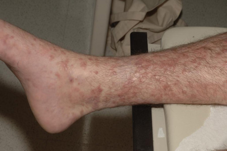
გამოკვლევა
კაპოშის სარკომის თავდაპირველი შეფასება საჭიროებს სრულ გასინჯვას. განსაკუთრებული ყურადღება ეთმობა ხშირად ჩართულ ადგილებს, როგორიცაა ქვედა კიდურები, სახე, პირის ღრუს ლორწოვანი გარსი და გენიტალიები.[4]Cesarman E, Damania B, Krown SE, et al. Kaposi sarcoma. Nat Rev Dis Primers. 2019 Jan 31;5(1):9.
https://www.nature.com/articles/s41572-019-0060-9
http://www.ncbi.nlm.nih.gov/pubmed/30705286?tool=bestpractice.com
[5]Esser S, Schöfer H, Hoffmann C, et al. S1 guidelines for the Kaposi sarcoma. J Dtsch Dermatol Ges. 2022 Jun;20(6):892-904.
https://onlinelibrary.wiley.com/doi/10.1111/ddg.14788
http://www.ncbi.nlm.nih.gov/pubmed/35657085?tool=bestpractice.com
ლიმფური კვანძები უნდა შემოწმდეს.[2]National Comprehensive Cancer Network. NCCN clinical practice guidelines in oncology: Kaposi sarcoma [internet publication].
https://www.nccn.org/guidelines/category_1
[5]Esser S, Schöfer H, Hoffmann C, et al. S1 guidelines for the Kaposi sarcoma. J Dtsch Dermatol Ges. 2022 Jun;20(6):892-904.
https://onlinelibrary.wiley.com/doi/10.1111/ddg.14788
http://www.ncbi.nlm.nih.gov/pubmed/35657085?tool=bestpractice.com
ლორწოვანი გარსის დაზიანებები და ლიმფედემა შეიძლება იყოს გავრცობილი და უნდა იყოს დოკუმენტირებული.
კაპოშის სარკომის დაზიანებას შემდეგი ტიპური მახასიათებლები აქვს:[3]Lebbe C, Garbe C, Stratigos AJ, et al; European Dermatology Forum (EDF), the European Association of Dermato-Oncology (EADO), and the European Organisation for Research and Treatment of Cancer (EORTC). Diagnosis and treatment of Kaposi's sarcoma: European consensus-based interdisciplinary guideline (EDF/EADO/EORTC). Eur J Cancer. 2019 Jun;114:117-27.
http://www.ncbi.nlm.nih.gov/pubmed/31096150?tool=bestpractice.com
მულტიფოკალური
ასიმეტრიულად განლაგებული
სხვადასხვა ზომის
სხვადასხვა ფერის (ვარდისფერი, წითელი, იასამნისფერი, ყავისფერი ან ლურჯი)
კვანძოვანი
პაპულური
ფოლაქისმაგვარი
ბულოზური
გამაგრებული (ხის მაგვარი)
მეჭეჭისებრი
სოკოსებრი ზრდის
წყლულოვანი
ინფიცირებული.
[Figure caption and citation for the preceding image starts]: მრავლობითი ვარდისფერი-იასამნისფერი კაპოშის სარკომის კვანძები ქვედა კიდურებზეFrom the collection of Dr Bruce J. Dezube; used with permission [Citation ends].[Figure caption and citation for the preceding image starts]: კაპოშის სარკომა, იასამნისფერი-მოყავისფრო ფოლაქი ტერფზეFrom the collection of Dr Bruce J. Dezube; used with permission [Citation ends].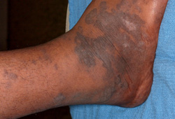
ვისცერული ჩართულობა, კანისა და ლორწოვანის დაზიანებების ფონზე ან მათ გარეშე, უნდა დადგინდეს ლიმფური კვანძების, გასტროინტესტინური ტრაქტისა და რესპირატორული ტრაქტის დაკვირვებით.
კვლევები
თუ აივ-ის სტატუსი უცნობია, აივ ტესტირება უნდა ჩაუტარდეს იმ პირებს, რომლებსაც აღენიშნებათ კაპოშის სარკომა (KS).[2]National Comprehensive Cancer Network. NCCN clinical practice guidelines in oncology: Kaposi sarcoma [internet publication].
https://www.nccn.org/guidelines/category_1
[3]Lebbe C, Garbe C, Stratigos AJ, et al; European Dermatology Forum (EDF), the European Association of Dermato-Oncology (EADO), and the European Organisation for Research and Treatment of Cancer (EORTC). Diagnosis and treatment of Kaposi's sarcoma: European consensus-based interdisciplinary guideline (EDF/EADO/EORTC). Eur J Cancer. 2019 Jun;114:117-27.
http://www.ncbi.nlm.nih.gov/pubmed/31096150?tool=bestpractice.com
[5]Esser S, Schöfer H, Hoffmann C, et al. S1 guidelines for the Kaposi sarcoma. J Dtsch Dermatol Ges. 2022 Jun;20(6):892-904.
https://onlinelibrary.wiley.com/doi/10.1111/ddg.14788
http://www.ncbi.nlm.nih.gov/pubmed/35657085?tool=bestpractice.com
თუ პაციენტი აივ დადებითია, უნდა ჩატარდეს CD4+ T-უჯრედების რაოდენობრივი ანალიზი და განისაზღვროს აივ ვირუსული დატვირთვა.
კლინიკურად საეჭვო დაზიანებები უნდა დადასტურდეს მცირე ზომის (2-4 მმ) პუნქციური ბიოფსიით და ჰისტოპათოლოგიით. კანის პუნქციური ბიოფსია ადვილად ტარდება და გვეხმარება დიფდიაგნოსტიკაში. ჰისტოპათოლოგიურად გამოვლინდება ატიპური თითისტარისებრი უჯრედები.[30]Radu O, Pantanowitz L. Kaposi sarcoma. Arch Pathol Lab Med. 2013 Feb;137(2):289-94.
https://meridian.allenpress.com/aplm/article/137/2/289/65260/Kaposi-Sarcoma
http://www.ncbi.nlm.nih.gov/pubmed/23368874?tool=bestpractice.com
იმუნოჰისტოქიმიური ტექნიკის გამოყენება შესაძლებელია კაპოშის სარკომით დაზიანებული უჯრედების მიერ ექსპრესირებული შემდეგი მარკერების გამოსავლენად: ლატენტურობასთან ასოცირებული ბირთვული ანტიგენ-1 (LANA-1, HHV-8-ის სუროგატი მარკერი); CD31/CD34 (სისხლძარღვთა ენდოთელური უჯრედების იმუნომარკერები) და D2-40 (ლიმფური უჯრედის იმუნომარკერი).[31]Jary A, Veyri M, Gothland A, et al. Kaposi's sarcoma-associated herpesvirus, the etiological agent of all epidemiological forms of Kaposi's sarcoma. Cancers (Basel). 2021 Dec 9;13(24):6208.
https://www.doi.org/10.3390/cancers13246208
http://www.ncbi.nlm.nih.gov/pubmed/34944828?tool=bestpractice.com
[32]Rosado FG, Itani DM, Coffin CM, et al. Utility of immunohistochemical staining with FLI1, D2-40, CD31, and CD34 in the diagnosis of acquired immunodeficiency syndrome-related and non-acquired immunodeficiency syndrome-related Kaposi sarcoma. Arch Pathol Lab Med. 2012 Mar;136(3):301-4.
https://meridian.allenpress.com/aplm/article/136/3/301/185684/Utility-of-Immunohistochemical-Staining-With-FLI1
http://www.ncbi.nlm.nih.gov/pubmed/22372906?tool=bestpractice.com
LANA-1-ის დადებითმა შედეგმა შეიძლება დაადასტუროს KS დიაგნოზი; CD31 და CD34 კვლევა შეიძლება სასარგებლო იყოს,თუ გაურკვეველია, არის თუ არა სიმსივნე სისხლძარღვოვანი წარმოშობის.[2]National Comprehensive Cancer Network. NCCN clinical practice guidelines in oncology: Kaposi sarcoma [internet publication].
https://www.nccn.org/guidelines/category_1
[5]Esser S, Schöfer H, Hoffmann C, et al. S1 guidelines for the Kaposi sarcoma. J Dtsch Dermatol Ges. 2022 Jun;20(6):892-904.
https://onlinelibrary.wiley.com/doi/10.1111/ddg.14788
http://www.ncbi.nlm.nih.gov/pubmed/35657085?tool=bestpractice.com
[32]Rosado FG, Itani DM, Coffin CM, et al. Utility of immunohistochemical staining with FLI1, D2-40, CD31, and CD34 in the diagnosis of acquired immunodeficiency syndrome-related and non-acquired immunodeficiency syndrome-related Kaposi sarcoma. Arch Pathol Lab Med. 2012 Mar;136(3):301-4.
https://meridian.allenpress.com/aplm/article/136/3/301/185684/Utility-of-Immunohistochemical-Staining-With-FLI1
http://www.ncbi.nlm.nih.gov/pubmed/22372906?tool=bestpractice.com
ადამიანის ჰერპესვირუსი-8-ის განსაზღვრა (პოლიმერაზული ჯაჭვური რეაქცია [PCR]) და HHV-8 ანტისხეულების ანალიზი, როგორც წესი, არ არის გამართლებული რუტინულ კლინიკურ პრაქტიკაში, მაგრამ შეიძლება ხელმისაწვდომი იყოს ინდივიდუალურ შემთხვევებში.[3]Lebbe C, Garbe C, Stratigos AJ, et al; European Dermatology Forum (EDF), the European Association of Dermato-Oncology (EADO), and the European Organisation for Research and Treatment of Cancer (EORTC). Diagnosis and treatment of Kaposi's sarcoma: European consensus-based interdisciplinary guideline (EDF/EADO/EORTC). Eur J Cancer. 2019 Jun;114:117-27.
http://www.ncbi.nlm.nih.gov/pubmed/31096150?tool=bestpractice.com
[5]Esser S, Schöfer H, Hoffmann C, et al. S1 guidelines for the Kaposi sarcoma. J Dtsch Dermatol Ges. 2022 Jun;20(6):892-904.
https://onlinelibrary.wiley.com/doi/10.1111/ddg.14788
http://www.ncbi.nlm.nih.gov/pubmed/35657085?tool=bestpractice.com
პაციენტებში, რომლებიც საჭიროებენ სისტემურ ქიმიოთერაპიას, უნდა შეფასდეს ძვლის ტვინის, ასევე ღვიძლისა და თირკმლის ფუნქციები. სისხლის საერთო ანალიზი (CBC) და სრული მეტაბოლური პანელი(სისხლის ბიოქიმიური ანალიზი) წარმოადგენს საწყისი ლაბორატორიული კვლევების შემადგენელს.[2]National Comprehensive Cancer Network. NCCN clinical practice guidelines in oncology: Kaposi sarcoma [internet publication].
https://www.nccn.org/guidelines/category_1
გამოსახულებითი და შემდგომი კვლევები
გულმკერდის რენტგენოგრაფიით ფილტვის კვლევა გვეხმარება რესპირატორული სიმპტომების მქონე პაციენტებში ფილტვის KS-ის სკრინინგში . [2]National Comprehensive Cancer Network. NCCN clinical practice guidelines in oncology: Kaposi sarcoma [internet publication].
https://www.nccn.org/guidelines/category_1
[5]Esser S, Schöfer H, Hoffmann C, et al. S1 guidelines for the Kaposi sarcoma. J Dtsch Dermatol Ges. 2022 Jun;20(6):892-904.
https://onlinelibrary.wiley.com/doi/10.1111/ddg.14788
http://www.ncbi.nlm.nih.gov/pubmed/35657085?tool=bestpractice.com
უნდა გამოირიცხოს ფილტვის თანმხლები ინფექცია. გულმკერდის რენტგენოლოგიური კვლევით ცვლილებების მქონე პაციენტებში, ფილტვის KS-ის დიაგნოზის დასადასტურებლად, შესაძლებელია გულმკერდის კომპიუტერული ტომოგრაფიის (CT) და ბრონქოსკოპიის ჩატარება გახდეს საჭირო.[2]National Comprehensive Cancer Network. NCCN clinical practice guidelines in oncology: Kaposi sarcoma [internet publication].
https://www.nccn.org/guidelines/category_1
[5]Esser S, Schöfer H, Hoffmann C, et al. S1 guidelines for the Kaposi sarcoma. J Dtsch Dermatol Ges. 2022 Jun;20(6):892-904.
https://onlinelibrary.wiley.com/doi/10.1111/ddg.14788
http://www.ncbi.nlm.nih.gov/pubmed/35657085?tool=bestpractice.com
სისხლდენის რისკის გამო, შედარებით უკუნაჩვენებია რესპირატორული ტრაქტის დაზიანებული კერების ბიოფსია.[33]Pitchenik AE, Fischl MA, Saldana MJ. Kaposi's sarcoma of the tracheobronchial tree. Clinical, bronchoscopic, and pathologic features. Chest. 1985 Jan;87(1):122-4.
http://www.ncbi.nlm.nih.gov/pubmed/3965256?tool=bestpractice.com
[34]Zibrak JD, Silvestri RC, Costello P, et al. Bronchoscopic and radiologic features of Kaposi's sarcoma involving the respiratory system. Chest. 1986 Oct;90(4):476-9.
http://www.ncbi.nlm.nih.gov/pubmed/3489584?tool=bestpractice.com
[35]Kodra A, Walczyszyn M, Grossman C, et al. Case report: pulmonary Kaposi sarcoma in a non-HIV patient. F1000Res. 2015 Oct 7;4:1013.
https://f1000research.com/articles/4-1013/v1
http://www.ncbi.nlm.nih.gov/pubmed/26664711?tool=bestpractice.com
ბიოფსია შეიძლება საჭირო გახდეს მასიური ლიმფადენოპათიის შემთხვევაში, დაზიანებების სხვა გენეზის გამოსარიცხად. თუ სავარაუდოა ვისცერალური ჩართულობა, განავალში ფარული სისხლდენის ტესტი გვეხმარება გასტროინტესტინური ტრაქტის დაავადებების გამოვლენაში.[2]National Comprehensive Cancer Network. NCCN clinical practice guidelines in oncology: Kaposi sarcoma [internet publication].
https://www.nccn.org/guidelines/category_1
კუჭ-ნაწლავის ტრაქტის ენდოსკოპია სიმპტომურ პირებში შეიძლება იყოს გამოსადეგი, მაგრამ არ არის რეკომენდებული რუტინული შეფასების ნაწილი რკინის დეფიციტის, კუჭ-ნაწლავიდან სისხლის დაკარგვის ან კუჭ-ნაწლავის ტრაქტის სიმპტომების არარსებობის შემთხვევაში.[2]National Comprehensive Cancer Network. NCCN clinical practice guidelines in oncology: Kaposi sarcoma [internet publication].
https://www.nccn.org/guidelines/category_1
[4]Cesarman E, Damania B, Krown SE, et al. Kaposi sarcoma. Nat Rev Dis Primers. 2019 Jan 31;5(1):9.
https://www.nature.com/articles/s41572-019-0060-9
http://www.ncbi.nlm.nih.gov/pubmed/30705286?tool=bestpractice.com
[29]Bower M, Palfreeman A, Alfa-Wali M, et al; British HIV Association. British HIV Association guidelines for HIV-associated malignancies 2014. HIV Med. 2014 Mar;15(Suppl 2):1-92.
https://onlinelibrary.wiley.com/doi/full/10.1111/hiv.12136
http://www.ncbi.nlm.nih.gov/pubmed/24528810?tool=bestpractice.com
კომპიუტერული ტომოგრაფია, პოზიტრონულ-ემისიური ტომოგრაფია (PET) და მაგნიტური რეზონანსის ტომოგრაფია (MRI) შეიძლება სასარგებლო იყოს ღრმა კვანძოვანი და ვისცერული კაპოშის სარკომის, ასევე ძვლის დაზიანებების შესაფასებლად. ძვლის დაზიანებები უკეთესად ვლინდება კომპიუტერული ტომოგრაფიისა და მაგნიტურ-რეზონანსული ტომოგრაფიის საშუალებით, რადგან ჩვეულებრივი რენტგენის ან ძვლის სკანირების დროს ისინი ხშირად შეუმჩნეველი რჩება .[36]Caponetti G, Dezube BJ, Restrepo C, et al. Kaposi sarcoma of the musculoskeletal system: a review of 66 patients. Cancer. 2007 Mar 15;109(6):1040-52.
https://acsjournals.onlinelibrary.wiley.com/doi/10.1002/cncr.22500
http://www.ncbi.nlm.nih.gov/pubmed/17265518?tool=bestpractice.com
თუ დაგეგმილია ქიმიოთერაპია ან სხივური თერაპია, რეპროდუქციული ასაკის პაციენტებში უნდა ჩატარდეს ორსულობის ტესტირება.[5]Esser S, Schöfer H, Hoffmann C, et al. S1 guidelines for the Kaposi sarcoma. J Dtsch Dermatol Ges. 2022 Jun;20(6):892-904.
https://onlinelibrary.wiley.com/doi/10.1111/ddg.14788
http://www.ncbi.nlm.nih.gov/pubmed/35657085?tool=bestpractice.com
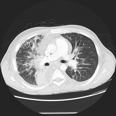
[Figure caption and citation for the preceding image starts]: კაპოშის სარკომა; ფოტომიკროგრაფიული ჰისტოპათოლოგია, ვლინდება სისხლძარღვოვანი, თითისტარისებრი სიმსივნური უჯრედების ფასციკულები (ჰემატოქსილინი და ეოზინი)From the collection of Dr Liron Pantanowitz; used with permission [Citation ends].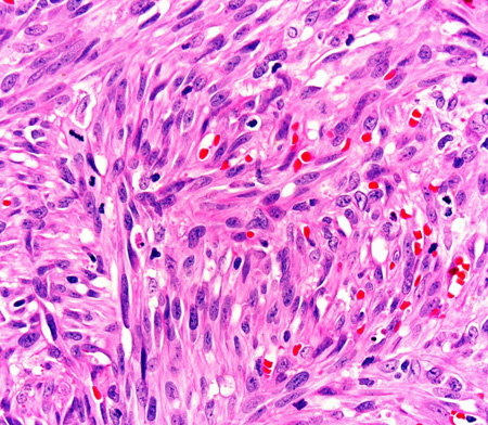 [Figure caption and citation for the preceding image starts]: გულმკერდის CT სკანირება, ვლინდება კაპოშის სარკომა ფილტვში, რეტიკულური სურათიFrom the collection of Dr Bruce J. Dezube; used with permission [Citation ends].