მიუხედავად იმისა, რომ მულტიფორმული ერითემა, როგორც წესი, მსუბუქი და თვითგანკურნებადი დაავადებაა, დეტალური ანამნეზი და კლინიკური გამოკვლევა დაგვეხმარება მაპროვოცირებელი ფაქტორების გამოვლენაში, რომელთაც პაციენტი მომავალში თავს აარიდებს.[41]Soares A, Sokumbi O. Recent updates in the treatment of erythema multiforme. Medicina (Kaunas). 2021 Sep 1;57(9):921.
https://www.ncbi.nlm.nih.gov/pmc/articles/PMC8467974
http://www.ncbi.nlm.nih.gov/pubmed/34577844?tool=bestpractice.com
ასევე საჭიროა გამოირიცხოს კანისა და ლორწოვანი გარსების უფრო მძიმე დაავადებები, როგორიცაა სტივენს-ჯონსონის სინდრომი (SJS) და ტოქსიკური ეპიდერმული ნეკროლიზი (TEN).[3]Grünwald P, Mockenhaupt M, Panzer R, et al. Erythema multiforme, Stevens-Johnson syndrome/toxic epidermal necrolysis - diagnosis and treatment. J Dtsch Dermatol Ges. 2020 Jun;18(6):547-53.
http://www.ncbi.nlm.nih.gov/pubmed/32469468?tool=bestpractice.com
მულტიფორმული ერითემის მსუბუქი ფორმის დროს მხოლოდ კანის გამოვლინებები გვხვდება, ხოლო მძიმე ფორმა მოიცავს ერთ ან მეტ ლორწოვან გარსსაც.
ორივე ფორმის შემთხვევაში მოცულია სხეულის სრული ზედაპირის <10%.
ანამნეზი.
აუცილებელია დეტალური ანამნეზის შეკრება ახლო წარსულში გადატანილ ინფექციასთან, განმეორებით დაავადებასა და ახალი მედიკამენტის ადმინისტრირებასთან დაკავშირებით. შესაძლო მიზეზების გულდასმით გამოკვლევა საჭიროა, სანამ შემთხვევას იდიოპათიურ ეტიოლოგიას მივაკუთვნებთ.[2]Sokumbi O, Wetter DA. Clinical features, diagnosis, and treatment of erythema multiforme: a review for the practicing dermatologist. Int J Dermatol. 2012 Aug;51(8):889-902.
http://onlinelibrary.wiley.com/doi/10.1111/j.1365-4632.2011.05348.x/full
http://www.ncbi.nlm.nih.gov/pubmed/22788803?tool=bestpractice.com
[41]Soares A, Sokumbi O. Recent updates in the treatment of erythema multiforme. Medicina (Kaunas). 2021 Sep 1;57(9):921.
https://www.ncbi.nlm.nih.gov/pmc/articles/PMC8467974
http://www.ncbi.nlm.nih.gov/pubmed/34577844?tool=bestpractice.com
ინფექციები: ყველაზე ხშირად დაკავშირებული ინფექციებია მარტივიჰერპესის ვირუსი და Mycoplasma pneumoniae-ს მიერ გამოწვეული ინფექციები.[39]Amode R, Ingen-Housz-Oro S, Ortonne N, et al. Clinical and histologic features of mycoplasma pneumoniae-related erythema multiforme: a single-center series of 33 cases compared with 100 cases induced by other causes. J Am Acad Dermatol. 2018 Jul;79(1):110-7.
http://www.ncbi.nlm.nih.gov/pubmed/29559400?tool=bestpractice.com
მულტიფორმული ერითემის შეტყობინებული სხვა მაპროვოცირებელი ინფექციური აგენტებია: ციტომეგალოვირუსი, ეპშტეინ-ბარის ვირუსი, SARS-CoV-2, В ჰეპატიტის ვირუსი, C ჰეპატიტის ვირუსი, გრიპის ვირუსი, აივ, ჰერპეს ზოსტერი, გარდნერელა, ჰისტოპლაზმოზი (თანმხლებ კვანძოვან ერითემასთან ერთად), კოქციდიოიდომიკოზი, კონტაგიოზური პუსტულური დერმატიტი (ცხვრებისა და თხების დაავადება, რომელიც გამოწვეულია პარაპოქს ვირუსით და შეიძლება გადაეცეს ადამიანებს) და სიფილისი.[2]Sokumbi O, Wetter DA. Clinical features, diagnosis, and treatment of erythema multiforme: a review for the practicing dermatologist. Int J Dermatol. 2012 Aug;51(8):889-902.
http://onlinelibrary.wiley.com/doi/10.1111/j.1365-4632.2011.05348.x/full
http://www.ncbi.nlm.nih.gov/pubmed/22788803?tool=bestpractice.com
[7]Ma JH, Smith S, Gordon LA. Acute HIV infection presenting as erythema multiforme in a 45-year-old heterosexual man. Med J Aust. 2015 Mar 16;202(5):273-5.
https://www.mja.com.au/journal/2015/202/5/acute-hiv-infection-presenting-erythema-multiforme-45-year-old-heterosexual-man
[10]Rossi L, Tiecco G, Venturini M, et al. Human orf with immune-mediated reactions: a systematic review. Microorganisms. 2023 Apr 27;11(5):1138.
https://www.ncbi.nlm.nih.gov/pmc/articles/PMC10224112
http://www.ncbi.nlm.nih.gov/pubmed/37317112?tool=bestpractice.com
[11]Kishore BN, Ankadavar NS, Kamath GH, et al. Varicella zoster with erythema multiforme in a young girl: a rare association. Indian J Dermatol. 2014 May;59(3):299-301.
http://www.e-ijd.org/article.asp?issn=0019-5154;year=2014;volume=59;issue=3;spage=299;epage=301;aulast=Kishore
http://www.ncbi.nlm.nih.gov/pubmed/24891667?tool=bestpractice.com
[12]Kasuya A, Sakabe J, Kageyama R, et al. Successful differentiation of herpes zoster-associated erythema multiforme from generalized extension of herpes by rapid polymerase chain reaction analysis. J Dermatol. 2014 Jun;41(6):542-4.
http://www.ncbi.nlm.nih.gov/pubmed/24909215?tool=bestpractice.com
[13]Olut AI, Erkek E, Ozunlu H, et al. Erythema multiforme associated with acute hepatitis B virus infection. Clin Exp Dermatol. 2006 Jan;31(1):137-8.[14]Saleh W, Alharbi H, Cha S. Increased prevalence of erythema multiforme in patients with COVID-19 infection or vaccination. Sci Rep. 2024 Feb 2;14(1):2801.
https://www.ncbi.nlm.nih.gov/pmc/articles/PMC10837137
http://www.ncbi.nlm.nih.gov/pubmed/38307870?tool=bestpractice.com
[15]Joseph RH, Haddad FA, Matthews AL, et al. Erythema multiforme after orf virus infection: a report of two cases and literature review. Epidemiol Infect. 2015 Jan;143(2):385-90.
https://www.cambridge.org/core/journals/epidemiology-and-infection/article/div-classtitleerythema-multiforme-after-orf-virus-infection-a-report-of-two-cases-and-literature-reviewdiv/324F9FDF41A60A2218CE2AEE7F78B25D/core-reader
http://www.ncbi.nlm.nih.gov/pubmed/24810660?tool=bestpractice.com
[16]Chiang MC, Chiang FC, Chang YT, et al. Erythema multiforme caused by treponema pallidum in a young patient with human immunodeficiency virus infection. J Clin Microbiol. 2010 Jul;48(7):2640-2.
https://jcm.asm.org/content/48/7/2640.long
http://www.ncbi.nlm.nih.gov/pubmed/20504989?tool=bestpractice.com
[37]Bitterman R, Oren I, Geffen Y, et al. Prolonged fever and splinter hemorrhages in an immunocompetent traveler with disseminated histoplasmosis. J Travel Med. 2013 Jan-Feb;20(1):57-9.
https://academic.oup.com/jtm/article/20/1/57/1817330
http://www.ncbi.nlm.nih.gov/pubmed/23279234?tool=bestpractice.com
[38]Bennardo L, Nisticò SP, Dastoli S, et al. Erythema multiforme and COVID-19: what do we know? Medicina (Kaunas). 2021 Aug 16;57(8):828.
https://www.ncbi.nlm.nih.gov/pmc/articles/PMC8401222
http://www.ncbi.nlm.nih.gov/pubmed/34441034?tool=bestpractice.com
მულტიფორმულ ერითემასთან დაკავშირებულია შემდეგი მედიკამენტები: ზოგიერთი ანტიბიოტიკი; დოცეტაქსელი ან პაკლიტაქსელი; იმუნური გამშვები მექანიზმის ინჰიბიტორები; სორაფენიბი; სიმსივნის ნეკროზის ფაქტორი (TNF) -ალფა ინჰიბიტორები; მალარიის საწინააღმდეგო პრეპარატები; ჰიდროქსიქლოროქინი; ლენალიდომიდი; მეთოტრექსატი; ანტიკონვულსანტები; სტატინები; ბისფოსფონატები; ანთების საწინააღმდეგო არასტეროიდული პრეპარატები (NSAIDs); მეტამიზოლი; ორალური კონტრაცეპტივები; იმიქიმოდი; ლიდოკაინი; ტრიკლოკარბანი; ბარიუმის კონტრასტი.[1]Lerch M, Mainetti C, Terziroli Beretta-Piccoli B, Harr T. Current perspectives on erythema multiforme. Clin Rev Allergy Immunol. 2018 Feb;54(1):177-84.
http://www.ncbi.nlm.nih.gov/pubmed/29352387?tool=bestpractice.com
[18]Borrás-Blasco J, Navarro-Ruiz A, Borrás C, et al. Adverse cutaneous reactions induced by TNF-alpha antagonist therapy. South Med J. 2009 Nov;102(11):1133-40.
http://www.ncbi.nlm.nih.gov/pubmed/19864977?tool=bestpractice.com
[19]Lenalidomide: Stevens-Johnson syndrome. Prescrire Int. 2010 Jun;19(107):125.
http://www.ncbi.nlm.nih.gov/pubmed/20740722?tool=bestpractice.com
[20]Ballester I, Guijarro J, Silvestre JF, et al. Erythema multiforme induced by imiquimod 5% cream. Int J Dermatol. 2014 Jul;53(7):e347-8.
http://www.ncbi.nlm.nih.gov/pubmed/24602041?tool=bestpractice.com
[21]Sai Keerthana PC, Anila KN, Reshma R. Naproxen induced erythema multiforme - a rare case report. Int J Pharm and Pharmaceutical Sci. 2017;9:294-5.
https://innovareacademics.in/journals/index.php/ijpps/article/viewFile/14903/9961
[22]Rodríguez-Pazos L, Sánchez-Aguilar D, Rodríguez-Granados MT, et al. Erythema multiforme photoinduced by statins. Photodermatol Photoimmunol Photomed. 2010 Aug;26(4):216-8.
http://www.ncbi.nlm.nih.gov/pubmed/20626826?tool=bestpractice.com
[23]Rodríguez-Pazos L, Gómez-Bernal S, Rodríguez-Granados MT, et al. Photodistributed erythema multiforme. Actas Dermosifiliogr. 2013 Oct;104(8):645-53.
https://www.actasdermo.org/en-photodistributed-erythema-multiforme-articulo-S1578219013001728
http://www.ncbi.nlm.nih.gov/pubmed/23962583?tool=bestpractice.com
[24]Utsunomiya A, Oyama N, Iino S, et al. A case of erythema multiforme major developed after sequential use of two immune checkpoint inhibitors, nivolumab and ipilimumab, for advanced melanoma: possible implication of synergistic and/or complementary immunomodulatory effects. Case Rep Dermatol. 2018 Jan 18;10(1):1-6.
https://www.ncbi.nlm.nih.gov/pmc/articles/PMC5836162
http://www.ncbi.nlm.nih.gov/pubmed/29515387?tool=bestpractice.com
[25]de Arruda JA, Silva P, Amaral MB, et al. Erythema multiforme induced by alendronate sodium in a geriatric patient: a case report and review of the literature. J Clin Exp Dent. 2017 Jul;9(7):e929-33.
https://www.ncbi.nlm.nih.gov/pmc/articles/PMC5549594
http://www.ncbi.nlm.nih.gov/pubmed/28828163?tool=bestpractice.com
[26]Abou Assalie N, Durcan R, Durcan L, et al. Hydroxychloroquine-induced erythema multiforme. J Clin Rheumatol. 2017 Mar;23(2):127-8.
https://www.ncbi.nlm.nih.gov/pmc/articles/PMC5321779
[27]Mantovani A, Álvares-Da-Silva MR. Anaphylaxis preceded by erythema multiforme with sorafenib: first case report. Ann Hepatol. 2019 Sep - Oct;18(5):777-9.
https://www.sciencedirect.com/science/article/pii/S1665268119300997?via%3Dihub
http://www.ncbi.nlm.nih.gov/pubmed/31085038?tool=bestpractice.com
თუმცა, ეს სია არ არის ამომწურავი და თქვენ უნდა შეამოწმოთ მედიკამენტის თაობაზე ინფორმაციის ადგილობრივი წყარო. ფენილბუტაზონის, ტრიკლოკარბანის, პაკლიტაქსელის და სტატინების ფონზე შესაძლოა განვითარდეს სინათლეზე ექსპოზიციის მიხედვით გავრცელებული დაზიანებები.[22]Rodríguez-Pazos L, Sánchez-Aguilar D, Rodríguez-Granados MT, et al. Erythema multiforme photoinduced by statins. Photodermatol Photoimmunol Photomed. 2010 Aug;26(4):216-8.
http://www.ncbi.nlm.nih.gov/pubmed/20626826?tool=bestpractice.com
[23]Rodríguez-Pazos L, Gómez-Bernal S, Rodríguez-Granados MT, et al. Photodistributed erythema multiforme. Actas Dermosifiliogr. 2013 Oct;104(8):645-53.
https://www.actasdermo.org/en-photodistributed-erythema-multiforme-articulo-S1578219013001728
http://www.ncbi.nlm.nih.gov/pubmed/23962583?tool=bestpractice.com
ვაქცინები და ალერგენები: დაავადებას შეიძლება ასევე იწვევდეს В ჰეპატიტის, ყვავილის, ჩუტყვავილას, მენინგოკოკის, ადამიანის პაპილომავირუსისა და SARS-CoV-2-ის საწინააღმდეგო ვაქცინები.[14]Saleh W, Alharbi H, Cha S. Increased prevalence of erythema multiforme in patients with COVID-19 infection or vaccination. Sci Rep. 2024 Feb 2;14(1):2801.
https://www.ncbi.nlm.nih.gov/pmc/articles/PMC10837137
http://www.ncbi.nlm.nih.gov/pubmed/38307870?tool=bestpractice.com
[28]Chahal D, Aleshin M, Turegano M, et al. Vaccine-induced toxic epidermal necrolysis: a case and systematic review. Dermatol Online J. 2018 Jan 15;24(1):13030/qt7qn5268s.
https://escholarship.org/uc/item/7qn5268s
http://www.ncbi.nlm.nih.gov/pubmed/29469759?tool=bestpractice.com
[40]Yousefian M, Khadivi A. Occurrence of erythema multiforme following COVID-19 vaccination: a review. Clin Exp Vaccine Res. 2023 Apr;12(2):87-96.
https://www.ncbi.nlm.nih.gov/pmc/articles/PMC10193109
http://www.ncbi.nlm.nih.gov/pubmed/37214146?tool=bestpractice.com
მულტიფორმული ერითემა ასევე შესაძლოა გამოიწვიოს კონტაქტურ ალერგენებზე (მაგ., როგორიცაა ტატუირება) ალერგიული პასუხმა.[29]Allione A, Dutto L, Castagna E, et al. Erythema multiforme caused by tattoo: a further case. Intern Emerg Med. 2011 Jun;6(3):263-5.
https://link.springer.com/article/10.1007%2Fs11739-010-0394-5
დაზიანებები, ტიპურ შემთხვევებში, მაპროვოცირებელი ფაქტორის მოქმედებიდან რამდენიმე დღის შემდეგ ვლინდება. ზოგიერთი დაზიანება თავდაპირველად სამიზნისმაგვარია; სხვა დაზიანებები იღებენ მცირე ზომის ერითემატოზული პაპულების ფორმას და შემდეგ გარდაიქმნებიან. დაზიანებები სწრაფად ვითარდება და 4-7 დღის განმავლობაში მათი რაოდენობა იმატებს. მდგომარეობამ შეიძლება გამოიწვიოს ზოგადი დისკომფორტი, თუმცა შეხორცების პერიოდამდე ქავილი არ ვლინდება. პირის ღრუს ლორწოვანი გარსის ჩართულობა შეიძლება განსაკუთრებით მტკივნეული იყოს პაციენტისთვის. უფრო მძიმე შემთხვევებში იზღუდება სითხეებისა და საკვების მიღება.
კლინიკური გამოკვლევა
მულტიფორმული ერითემა ვლინდება ტიპური სამიზნისმაგვარი დაზიანებებით (რგოლის ფორმის ერითემატოზული ბეჭდები, გარე ერითემატოზული ზონითა და ცენტრალური ბუშტუკით, რომელთა შორისაც კანი ნორმალური შეფერილობისაა) და ატიპური ტარგეტოიდული პაპულები (ცენტრალური ბუშტუკების გარეშე). დაზიანებების კლინიკური განვითარება ყველაზე მნიშვნელოვანი დიაგნოსტიკური ინსტრუმენტია, რადგან დამახასიათებელი სამიზნისმაგვარი დაზიანებები ხშირად კიდურებზე, სიმეტრიული განლაგებით ვლინდება. ტარგეტოიდული დაზიანებები უფრო ხშირად ცენტრიპეტალურად ვლინდება.[2]Sokumbi O, Wetter DA. Clinical features, diagnosis, and treatment of erythema multiforme: a review for the practicing dermatologist. Int J Dermatol. 2012 Aug;51(8):889-902.
http://onlinelibrary.wiley.com/doi/10.1111/j.1365-4632.2011.05348.x/full
http://www.ncbi.nlm.nih.gov/pubmed/22788803?tool=bestpractice.com
[3]Grünwald P, Mockenhaupt M, Panzer R, et al. Erythema multiforme, Stevens-Johnson syndrome/toxic epidermal necrolysis - diagnosis and treatment. J Dtsch Dermatol Ges. 2020 Jun;18(6):547-53.
http://www.ncbi.nlm.nih.gov/pubmed/32469468?tool=bestpractice.com
[4]Bastuji-Garin S, Rzany B, Stern RS, et al. Clinical classification of cases of toxic epidermal necrolysis, Stevens-Johnson syndrome, and erythema multiforme. Arch Dermatol. 1993 Jan;129(1):92-6.
http://www.ncbi.nlm.nih.gov/pubmed/8420497?tool=bestpractice.com
[5]Assier H, Bastuji-Garin S, Revuz J, et al. Erythema multiforme with mucous membrane involvement and Stevens-Johnson syndrome are clinically different disorders with distinct causes. Arch Dermatol. 1995 May;131(5):539-43.
http://www.ncbi.nlm.nih.gov/pubmed/7741539?tool=bestpractice.com
სამიზნე დაზიანებებთან ერთად სწრაფად განვითარებული სამიზნისმაგვარი ერთეულების არსებობა კიდევ უფრო ამყარებს მულტიფორმული ერითემის დიაგნოზს. პირის ღრუს, თვალების, ცხვირისა და გენიტალიების ლორწოვანი გარსის მემბრანები ასევე უნდა გამოვიკვლიოთ ეროზიების აღმოსაჩენად, რომლებიც ვლინდება მულტიფორმული ერითემის მძიმე ფორმის დროს. ზოგიერთი დამახასიათებელი სამიზნე დაზიანება და ლორწოვანის მინიმალური ჩართულობა მიუთითებს მულტიფორმულ ერითემაზე, განსაკუთრებით, მარტივი ჰერპესის ან Mycoplasma pneumoniae-ს ინფექციის ფონზე.
საჭიროა უფრო ფართო გასინჯვა რაიმე შესაძლო ინფექციური მიზეზის საპოვნელად. მარტივი ჰერპესის ვირუსით გამოწვეული ინფექცია ხასიათდება დაჯგუფებული ვეზიკულებით, რომლებიც ერითემატოზულ ფონზე ჩნდებიან. წითელი ფერის დაფის აპკი მიუთითებს Mycoplasma pneumoniae-ზე, ისევე როგორც ხიხინი და/ან მსტვინავი სუნთქვა.[Figure caption and citation for the preceding image starts]: სამიზნისმაგვარი დაზიანებები ხელისგულზეNanette Silverberg, MD-ის პირადი კოლექციიდან, გამოყენებულია ნებართვით [Citation ends].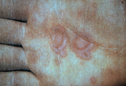 [Figure caption and citation for the preceding image starts]: სამიზნისმაგვარი და ტარგეტოიდული დაზიანებებიNanette Silverberg, MD-ის პირადი კოლექციიდან, გამოყენებულია ნებართვით [Citation ends].
 [Figure caption and citation for the preceding image starts]: სამიზნისმაგვარი დაზიანებები სახეზე და ლორწოვანი ეროზიები ქერქებით, მარტივი ჰერპესვირუსის განმეორებითი ეპიზოდიNanette Silverberg, MD-ის პირადი კოლექციიდან, გამოყენებულია ნებართვით [Citation ends].
ლაბორატორიიული კვლევები
მულტიფორმული ერითემის შემთხვევათა უმეტესობის დიაგნოსტირება შესაძლებელია მხოლოდ ანამნეზითა და კლინიკური გასინჯვით და შემდგომ გამოკვლევებს არ საჭიროებს. თუმცა, როცა კლინიკური გასინჯვის შემდეგ დიაგნოზი გაურკვეველია, შეიძლება ჩატარდეს ბიოფსია და ნიმუშის გამოკვლევა ჰემატოქსილინითა და ეოზინით. თუ ბიოფსიის შედეგი დამაჯერებელი არ არის, შესაძლებელია ასევე ბიოპტატის შეფასება იმუნოფლუორესცენტული კვლევით.
თუ კლინიკური გასინჯვით მულტიფორმული ერითემის მიზეზი არ ვლინდება, აღნიშნულის დასადგენად ტარდება ლაბორატორიული ტესტები. რადგანაც მულტიფორმულ ერითემას ინფექციებიდან ყველაზე ხშირად მარტივი ჰერპესის ვირუსი და მიკოპლაზმა იწვევს, საწყისი კვლევებია: სისხლის საერთო ანალიზი, ელექტროლიტები, მარტივი ჰერპესის ვირუსის სეროლოგია, ცივი აგლუტინინები, M pneumoniae-ს ტიტრი და/ან გულმკერდის რენტგენოგრაფია (დამოკიდებულია პაციენტის კლინიკურ მდგომარეობაზე). თუ აღნიშნული ტესტები უარყოფითია, შედარებით იშვიათი ინფექციური გამომწვევების გამოსარიცხად ტარდება სხვა კვლევები. ჰერპეს ზოსტერთან დაკავშირებული მულტიფორმული ერითემის გენერალიზებული ჰერპესისგან დიფერენცირება შესაძლებელია სწრაფი პოლიმერაზული ჯაჭვური რეაქციის მეშვეობით.[12]Kasuya A, Sakabe J, Kageyama R, et al. Successful differentiation of herpes zoster-associated erythema multiforme from generalized extension of herpes by rapid polymerase chain reaction analysis. J Dermatol. 2014 Jun;41(6):542-4.
http://www.ncbi.nlm.nih.gov/pubmed/24909215?tool=bestpractice.com
მარტივი ჰერპესის ვირუსის სეროლოგია შეიძლება გამოგვადგეს იმ შემთხვევაში, თუ მულტიფორმული ერითემა რამდენჯერმე განმეორდა, მაგრამ სპეციფიკური ჰერპესული დაზიანებები არ დაფიქსირებულა. ანტიდესმოპლაკინის ანტისხეულები შემჩნეულია განმეორებითი მულტიფორმული ერითემის მქონე პაციენტებში.[36]Wetter DA, Davis MD. Recurrent erythema multiforme: clinical characteristics, etiologic associations, and treatment in a series of 48 patients at Mayo Clinic, 2000 to 2007. J Am Acad Dermatol. 2010 Jan;62(1):45-53.
http://www.jaad.org/article/S0190-9622(09)00778-6/fulltext
http://www.ncbi.nlm.nih.gov/pubmed/19665257?tool=bestpractice.com
სტივენს-ჯონსონის სინდრომისა და ტოქსიკური ეპიდერმული ნეკროლიზის განსხვავება მულტიფორმული ერითემისაგან
უნდა გამოირიცხოს სხვა უფრო მძიმე რეაქციული პროცესები, მათ შორის სტივენს-ჯონსონის სინდრომი და ტოქსიკური ეპიდერმული ნეკროლიზი. საკვების მიღების შეზღუდვა და ტკივილი მოშარდვისას უფრო ხშირია ამ დაავადებების დროს, თუმცა შეიძლება შეგვხვდეს მულტიფორმული ერითემის მძიმე ფორმასთან ერთადაც.[2]Sokumbi O, Wetter DA. Clinical features, diagnosis, and treatment of erythema multiforme: a review for the practicing dermatologist. Int J Dermatol. 2012 Aug;51(8):889-902.
http://onlinelibrary.wiley.com/doi/10.1111/j.1365-4632.2011.05348.x/full
http://www.ncbi.nlm.nih.gov/pubmed/22788803?tool=bestpractice.com
[3]Grünwald P, Mockenhaupt M, Panzer R, et al. Erythema multiforme, Stevens-Johnson syndrome/toxic epidermal necrolysis - diagnosis and treatment. J Dtsch Dermatol Ges. 2020 Jun;18(6):547-53.
http://www.ncbi.nlm.nih.gov/pubmed/32469468?tool=bestpractice.com
[4]Bastuji-Garin S, Rzany B, Stern RS, et al. Clinical classification of cases of toxic epidermal necrolysis, Stevens-Johnson syndrome, and erythema multiforme. Arch Dermatol. 1993 Jan;129(1):92-6.
http://www.ncbi.nlm.nih.gov/pubmed/8420497?tool=bestpractice.com
[5]Assier H, Bastuji-Garin S, Revuz J, et al. Erythema multiforme with mucous membrane involvement and Stevens-Johnson syndrome are clinically different disorders with distinct causes. Arch Dermatol. 1995 May;131(5):539-43.
http://www.ncbi.nlm.nih.gov/pubmed/7741539?tool=bestpractice.com
[9]Auquier-Dunant A, Mockenhaupt M, Maldi L, et al. Correlations between clinical patterns and causes of erythema multiforme majus, Stevens-Johnson syndrome, and toxic epidermal necrolysis: results of an international prospective study. Arch Dermatol. 2002 Aug;138(8):1019-24.
https://jamanetwork.com/journals/jamadermatology/fullarticle/478935
http://www.ncbi.nlm.nih.gov/pubmed/12164739?tool=bestpractice.com
[42]Cote B, Wechsler J, Bastuji-Garin S. Clinicopathologic correlation in erythema multiforme and Stevens-Johnson syndrome. Arch Dermatol. 1995 Nov;131(11):1268-72.
http://www.ncbi.nlm.nih.gov/pubmed/7503570?tool=bestpractice.com
სტივენს-ჯონსონის სინდრომი მოიცავს სხეულის სრული ზედაპირის <10% ნაწილს, უფრო ხშირად ჩართულია პირის ღრუ და გენიტალიების ლორწოვანი გარსები. ხშირად შესაძლებელია "დამნაშავე" მედიკამენტის იდენტიფიცირება. ტოქსიკური ეპიდერმული ნეკროლიზი იწვევს კანის ექტენსიურ აშრევებას, როგორც წესი, სხეულის სრული ზედაპირის >30%-ს მოიცავს. თუ რთულია მულტიფორმული ერითემის შესაძლო შემთხვევის განსხვავება აღნიშნული ორი პათოლოგიისაგან, შესაძლებელია 2 ტესტის ჩატარება:
ტოქსიკური ეპიდერმული ნეკროლიზის დროს გვხვდება ასბო-ჰანსენის ნიშანი - ლატერალური ზეწოლით ბუშტუკები ფიზიკურად დიდდება, რაც გამოხატავს ბაზალური კერატინოციტების ნეკროზს. სტივენს-ჯონსონის სინდრომისა და ტოქსიკური ეპიდერმული ნეკროლიზის დროს ასევე აღინიშნება ნიკოლსკის ნიშანი- კანი აშრევდება შეხებისას. ეს ნიშანი მულტიფორმული ერითემისთვის არ არის დამახასიათებელი.[2]Sokumbi O, Wetter DA. Clinical features, diagnosis, and treatment of erythema multiforme: a review for the practicing dermatologist. Int J Dermatol. 2012 Aug;51(8):889-902.
http://onlinelibrary.wiley.com/doi/10.1111/j.1365-4632.2011.05348.x/full
http://www.ncbi.nlm.nih.gov/pubmed/22788803?tool=bestpractice.com
ბიოფსია და ახალი გაყინული ქსოვილის შეფასება შეიძლება დაგვეხმაროს ნეკროზული კერატინოციტების გამოვლენაში სტივენს-ჯონსონის სინდრომისა და ტოქსიკური ეპიდერმული ნეკროლიზის დროს. მულტიფორმული ერითემის ჰისტოპათოლოგიური სურათი უფრო ხშირად მოიცავს ერითროციტებისა და მონოციტების ინფილტრატებს; არ ვლინდება ეპიდერმისის ნეკროზი და ანთებითი ინფილტრატი დერმისში.[2]Sokumbi O, Wetter DA. Clinical features, diagnosis, and treatment of erythema multiforme: a review for the practicing dermatologist. Int J Dermatol. 2012 Aug;51(8):889-902.
http://onlinelibrary.wiley.com/doi/10.1111/j.1365-4632.2011.05348.x/full
http://www.ncbi.nlm.nih.gov/pubmed/22788803?tool=bestpractice.com
[3]Grünwald P, Mockenhaupt M, Panzer R, et al. Erythema multiforme, Stevens-Johnson syndrome/toxic epidermal necrolysis - diagnosis and treatment. J Dtsch Dermatol Ges. 2020 Jun;18(6):547-53.
http://www.ncbi.nlm.nih.gov/pubmed/32469468?tool=bestpractice.com
[4]Bastuji-Garin S, Rzany B, Stern RS, et al. Clinical classification of cases of toxic epidermal necrolysis, Stevens-Johnson syndrome, and erythema multiforme. Arch Dermatol. 1993 Jan;129(1):92-6.
http://www.ncbi.nlm.nih.gov/pubmed/8420497?tool=bestpractice.com
[5]Assier H, Bastuji-Garin S, Revuz J, et al. Erythema multiforme with mucous membrane involvement and Stevens-Johnson syndrome are clinically different disorders with distinct causes. Arch Dermatol. 1995 May;131(5):539-43.
http://www.ncbi.nlm.nih.gov/pubmed/7741539?tool=bestpractice.com
[32]Sundram U. A review of important skin disorders occurring in the posttransplantation patient. Adv Anat Pathol. 2014 Sep;21(5):321-9.
http://www.ncbi.nlm.nih.gov/pubmed/25105934?tool=bestpractice.com
[42]Cote B, Wechsler J, Bastuji-Garin S. Clinicopathologic correlation in erythema multiforme and Stevens-Johnson syndrome. Arch Dermatol. 1995 Nov;131(11):1268-72.
http://www.ncbi.nlm.nih.gov/pubmed/7503570?tool=bestpractice.com
იხილეთ სტივენს-ჯონსონის სინდრომი და ტოქსიკური ეპიდერმული ნეკროლიზი (დიაგნოსტიკური მიდგომა).