Investigations
1st investigations to order
liver function tests
Test
Aminotransferase (aspartate aminotransferase [AST] and alanine aminotransferase [ALT]) levels increase with hepatocellular damage. Normal AST and ALT levels do not preclude the diagnosis of cirrhosis.[41] Aminotransferase levels bear little to no relationship to frequency of complications or death.[42]
ALT levels are greater than those of AST in most chronic liver diseases (except for alcohol-related liver disease), but this finding may be reversed with progression of liver disease. An AST/ALT ratio of ≥1 is thought to be a predictor of cirrhosis.[43]
Alkaline phosphatase and gamma-glutamyl transferase (GGT) levels increase in cholestasis (resulting from primary biliary cholangitis and primary sclerosing cholangitis), with minimal derangement of AST and ALT.
Total bilirubin may be normal in patients with compensated cirrhosis, but as the cirrhosis progresses, serum levels generally rise.
Result
usually deranged
gamma-glutamyl transferase (GGT)
Test
Increase in this liver microsomal enzyme represents enzyme activation, which can be induced by alcohol and certain drugs and is also observed in metabolic dysfunction-associated steatotic liver disease (MASLD).
Increased in cholestasis along with alkaline phosphatase (ALP).
GGT is not significantly present in bone, such that concomitant elevated GGT and ALP indicate the liver as the source of the ALP.
Result
elevated
serum albumin
Test
A decrease in serum albumin is a marker of hepatic synthetic dysfunction.
Result
reduced
serum sodium
Test
Hyponatraemia is a common finding in patients with cirrhosis with associated ascites, and worsens as the liver disease progresses.
Result
reduced
serum potassium
prothrombin time
Test
Prolongation of the prothrombin time is a marker of hepatic synthetic dysfunction.
Result
prolonged
platelet count
Test
The presence of thrombocytopenia (platelet count <150,000/microlitre) is the most sensitive and specific laboratory finding for the diagnosis of cirrhosis in the setting of chronic liver disease and results from portal hypertension with hypersplenism and platelet sequestration.[48]
Result
reduced
antibodies to hepatitis C virus
Test
Presence of immunoglobulin G (IgG) antibodies to hepatitis C virus (confirmed with hepatitis C virus-RNA) is indicative of chronic hepatitis C infection.[49]
Result
present if patient has chronic hepatitis C infection
hepatitis B surface antigen ± hepatitis B DNA assay
Test
A detectable HBsAg or viraemia on a highly sensitive hepatitis B DNA assay indicates chronic hepatitis B infection.[49]
Hepatitis B e-antigen/e-antibody, genotype, and viral load should be measured to assess disease phase and guide further treatment.
Result
HBsAg present or hepatitis B viraemia detected if patient has hepatitis B infection
Investigations to consider
total iron, total iron binding capacity (TIBC), transferrin saturation, and serum ferritin
Test
The initial screening tests for haemochromatosis are total iron and total iron binding capacity, in order to calculate the transferrin saturation (iron/TIBC), and serum ferritin.
If the transferrin saturation is elevated (>45%), further testing with ferritin and possible genetic testing (C282Y and H63D mutation analysis) should be undertaken.
If the transferrin saturation and ferritin are elevated, HFE genotyping is performed.[50]
Result
elevated transferrin saturation and elevated ferritin in haemochromatosis
antinuclear antibody
Test
Tests for autoimmune hepatitis
Result
present in autoimmune hepatitis
antismooth muscle antibody
Test
Result
present in autoimmune hepatitis
liver kidney microsomal antibody
Test
Liver kidney microsomal antigen antibodies (anti-LSM) are autoantibodies that target the CYP2D6 enzyme which is primarily found in the liver cells. Development of anti-LSM antibodies is associated with autoimmune hepatitis.
Result
present in autoimmune hepatitis
antimitochondrial antibody
Test
Specifically the M2 antibody.
Result
present in primary biliary cholangitis
serum immunoglobulins
Test
IgA, IgG, and IgM levels should be evaluated.
Result
frequently elevated in patients with cirrhosis
serum ceruloplasmin
Test
Result
low in Wilson's disease
plasma alpha-1 antitrypsin
Test
Approximately 15% of adults with alpha-1 antitrypsin deficiency develop cirrhosis.[52]
Plasma alpha-1 antitrypsin levels are used as a screening test.
Alpha-1 antitrypsin is an acute phase protein and may be elevated in inflammation.
Further testing can be done to confirm the diagnosis with protein electrophoresis and phenotyping.
Result
reduced in alpha-1 antitrypsin deficiency
alpha-fetoprotein
Test
The finding of elevated alpha-fetoprotein in a patient with cirrhosis should raise concern for the development of hepatocellular carcinoma.[53] However, this tumour marker may also be elevated on the basis of chronic liver disease and inflammation in the absence of hepatocellular carcinoma; therefore, cross-sectional imaging is indicated to exclude the presence of hepatic lesions.
Result
normal or raised
abdominal ultrasound
Test
Signs of advanced cirrhosis may be detected using abdominal ultrasound.
Signs of portal hypertension: ascites, splenomegaly, increased diameter of the portal vein (≥13 mm), or collateral vessels.
In combination with a strong clinical suspicion, the above findings suffice for the diagnosis of cirrhosis without the need for a confirmatory liver biopsy.
A normal abdominal ultrasound does not exclude significant liver disease.
Result
liver surface nodularity, small liver, possible hypertrophy of left/caudate lobe, ascites, splenomegaly, increased diameter of the portal vein (≥13 mm), or collateral vessels
abdominal CT
Test
Signs of advanced cirrhosis may be detected using abdominal cross-sectional imaging.
Signs of portal hypertension: ascites, splenomegaly, collateral circulation.
In combination with a strong clinical suspicion, the above findings suffice for the diagnosis of cirrhosis without the need of a confirmatory liver biopsy.
Result
liver surface nodularity, small liver, possible hypertrophy of left/caudate lobe, evidence of ascites, or collateral circulation
abdominal MRI
Test
Signs of advanced cirrhosis may be detected using MRI of the liver.
Signs of portal hypertension: ascites, splenomegaly, collateral circulation.
In combination with a strong clinical suspicion, the above findings suffice for the diagnosis of cirrhosis without the need of a confirmatory liver biopsy.
Result
liver surface nodularity, small liver, possible hypertrophy of left/caudate lobe, evidence of ascites, or collateral circulation
upper gastrointestinal endoscopy
Test
Identifies the presence of gastro-oesophageal varices or portal hypertensive gastropathy secondary to portal hypertension in patients with chronic liver disease, thus aiding the diagnosis of cirrhosis.
The Baveno VII criteria have been validated in several patient cohorts (with compensated advanced chronic liver disease) and suggest that screening endoscopy could be reserved for specific patients, based on liver stiffness measurement (LSM) and platelet count assessment.[54] In particular, patients with compensated cirrhosis, who have a liver stiffness <20 kPa (as measured by transient elastography) and platelet count >150 × 10⁹ cells/L, have a very low risk of having varices requiring treatment and can avoid screening endoscopy.[54] In practice, however, many centres still offer baseline endoscopy to all patients with liver cirrhosis.
In compensated patients with no or small varices at screening endoscopy, expanded Baveno VI criteria recommend screening for gastro-oesophageal varices at 1- to 3-year intervals thereafter.[87]
Baveno VII consensus guidelines conclude that patients who avoid screening endoscopy can be followed up by yearly repetition of transient elastography and platelet count. If LSM increases (≥20 kPa) or platelet count declines (≤150 × 10⁹/L), these patients should undergo screening endoscopy.[54]
[Figure caption and citation for the preceding image starts]: Oesophageal varices in a patient with portal hypertensionFrom the collection of Douglas G. Adler, MD [Citation ends].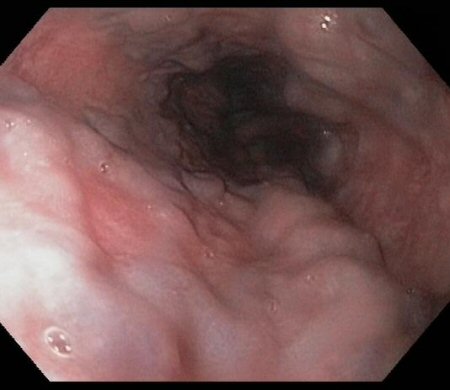
Result
gastro-oesophageal varices, portal hypertensive gastropathy
liver biopsy
Test
Liver biopsy remains the most specific and sensitive test for the diagnosis of cirrhosis. However, it is not necessary in patients with advanced liver disease and typical clinical, laboratory, and/or radiological findings of cirrhosis unless there is a need to determine the degree of inflammation.
In addition to confirming the diagnosis, liver biopsy may help to determine the aetiology of the underlying liver disease, although this is not always possible as characteristic features of the primary insult (e.g., metabolic dysfunction-associated steatotic liver disease or autoimmune hepatitis) may no longer be detectable by the time the procedure is carried out.
Liver biopsy is associated with risk of bleeding, perforation, and pneumothorax, among other complications.[63]
Result
architectural distortion of the liver parenchyma with formation of regenerative nodules
imaging-based non-invasive tests
Test
The American Association for the Study of Liver Diseases (AASLD) suggests a combination of imaging-based and blood-based techniques to detect significant fibrosis and advanced fibrosis, particularly in those undergoing initial fibrosis staining.[69] Imaging-based tests may be preferentially incorporated into the initial fibrosis staging process owing to their higher accuracy over blood-based techniques. These tests are recommended for the identification of significant fibrosis, advanced fibrosis, and cirrhosis in adults with chronic hepatitis B and hepatitis C infections and in those with metabolic dysfunction-associated steatotic liver disease (MASLD).[69] Imaging-based non-invasive testing may also be used in adults with alcohol-related liver disease or chronic cholestatic liver disease to detect advanced fibrosis or cirrhosis. Either transient elastography or magnetic resonance elastography is recommended by the AASLD to stage fibrosis in adults with chronic liver disease.[69] The AASLD advises against using imaging-based tests as a standalone test to assess regression or progression of liver fibrosis.[69] Ultrasound-based transient elastography is a useful tool for detecting hepatic fibrosis and cirrhosis without the need for liver biopsy. The Society of Radiologists in Ultrasound recommends a low cut-off value to exclude significant fibrosis, and a high cut-off value to indicate compensated advanced chronic liver disease.[70] Meta-analyses and prospective studies of transient elastography report excellent diagnostic accuracy for the diagnosis of cirrhosis (independent of the underlying disease) and the identification of fibrosis in patients with recurrent hepatitis C infection after liver transplantation.[71][72][73][74][75][76][77][78][79]
As with transient elastography, acoustic radiation force impulse (ARFI) imaging employs ultrasound to perform elastography. Magnetic resonance elastography is effective in measuring fibrosis, but its application is limited by cost, and it may not be possible if older metal prostheses are present in the patient. Staging of fibrosis may be possible using gadoxetic acid-enhanced MRI.[80] FibroScan® is a non-invasive modality that helps quantify and stage hepatic steatosis and fibrosis (especially advanced fibrosis and cirrhosis) by measuring the degree of liver stiffness using vibration-controlled transient elastography.[81][82] It can distinguish normal liver or minimal fibrosis from cirrhotic livers.[82] FibroScan® can be used as an option for the assessment of fibrosis or cirrhosis outside secondary and specialist care as it has a potential to detect liver disease earlier. It can be used in accordance with the national guidelines. The test should be recommended if it benefits people who lack adequate healthcare access (such as disabled people, people living in rural areas, or people from lower socio-economic backgrounds) and should be performed by trained operators.[82]
Result
staging of fibrosis
blood-based non-invasive tests
Test
The American Association for the Study of Liver Diseases (AASLD) suggests a combination of imaging-based and blood-based techniques to detect significant fibrosis and advanced fibrosis, particularly in those undergoing initial fibrosis staining.[69] Several fibrosis markers have been assessed. Some of these use combinations of routinely collected blood tests (e.g., non-alcoholic fatty liver disease [NAFLD] fibrosis score, fibrosis-4 [FIB-4], AST to platelet ratio index [APRI], AST/ALT ratio). [ NAFLD Fibrosis Score Opens in new window ] Others use specific molecular markers of fibrogenesis (e.g., enhanced liver fibrosis [ELF]). The utility of these markers is predominantly for excluding severe fibrosis, with normal values being reassuring. They do not perform well at differentiating intermediate stages of fibrosis. The AASLD recommends initial testing with APRI or FIB-4 markers to detect significant fibrosis, advanced fibrosis, or cirrhosis in adults with chronic hepatitis B and hepatitis C infections undergoing fibrosis staging prior to antiviral therapy.[83] The European Association for the Study of the Liver recommends that among non-invasive methods, the use of transient elastography has been mostly studied and seems to offer a higher diagnostic accuracy for the detection of cirrhosis in patients with chronic hepatitis B.[84]
Result
staging of fibrosis
portal pressure assessment
Test
The gold standard method to assess portal pressure in patients with cirrhosis is hepatic venous pressure gradient.[2] Calculating hepatic venous pressure gradient is an invasive procedure and, as a result, it is not routinely used in all patients with liver cirrhosis and portal hypertension.
Non-invasive assessment of clinically significant portal hypertension may be performed using a combination of liver stiffness measurement (LSM) and platelet count.[2][85] An LSM of ≥20 kPa suggests clinically significant portal hypertension.[86] Clinically significant portal hypertension is present in all patients with varices.
Result
LSM of ≥20 kPa suggests clinically significant portal hypertension
Use of this content is subject to our disclaimer