Images and videos
Images


Cirrhosis
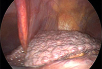
Oesophageal varices in a patient with portal hypertension
From the collection of Douglas G. Adler, MD
See this image in context in the following section/s:
Cirrhosis
Preoperative view of a small finger flexion contracture
From the collection of Dr C.M. Rodner; used with permission
See this image in context in the following section/s:

Cirrhosis
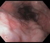
Laparoscopic view of a cirrhotic liver
Courtesy of Dr Eugene Schiff and Dr Lennox Jeffers; used with permission
See this image in context in the following section/s:
Cirrhosis
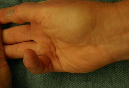
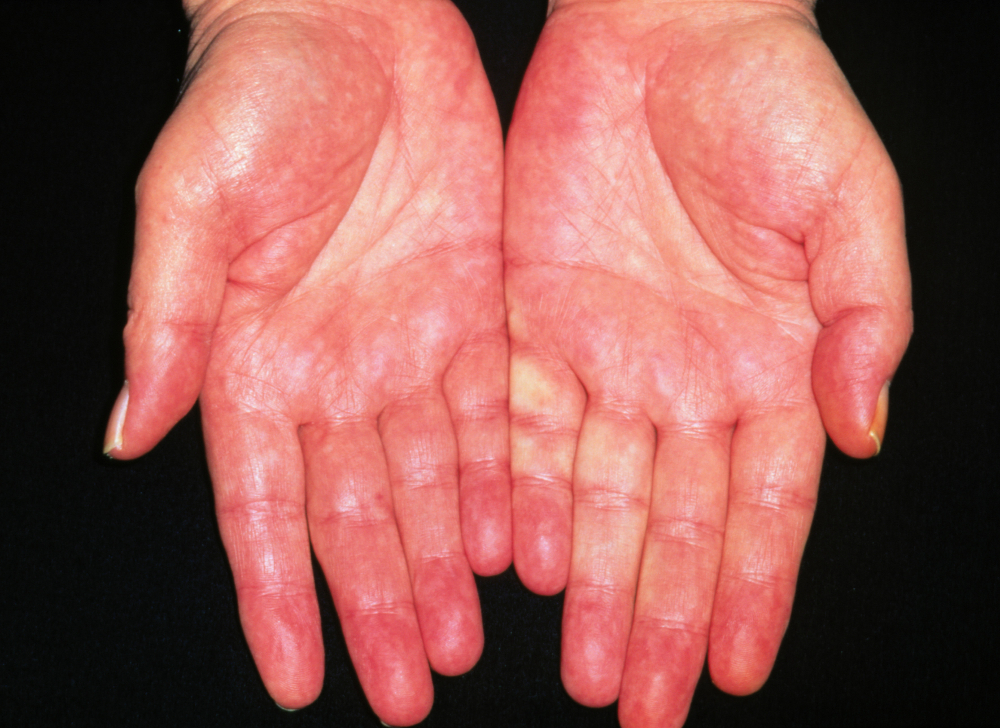
Liver palms erythema of adult alcoholic
Dr P. Marazzi / Science Photo Library; used with permission
See this image in context in the following section/s:
Cirrhosis
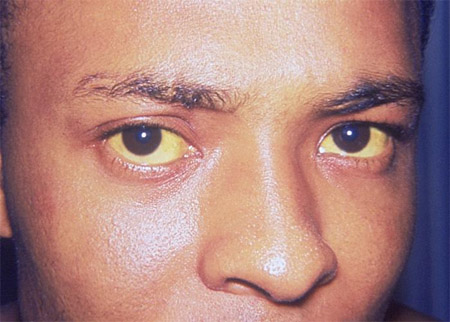
Icterus or jaundice
CDC. Dr Thomas F. Sellers/Emory University; used with permission
See this image in context in the following section/s:

Cirrhosis
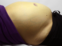
Ascites. View of the abdomen of a female patient with alcohol-related liver disease and cirrhosis, showing swelling due to ascites (accumulation of fluid in the peritoneal cavity), jaundice (yellowing of the skin), and bruising
Science Photo Library; used with permission
See this image in context in the following section/s:
Cirrhosis
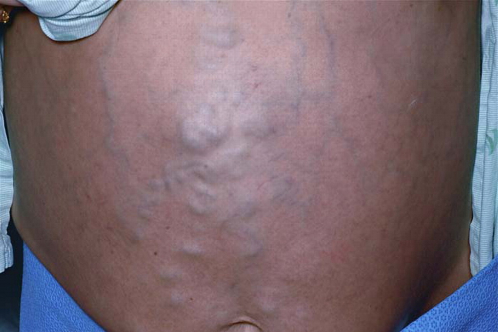
Caput medusa: dilated superficial (superior and inferior) epigastric veins radiating from a central large venous varix
Singh NK, Cheema U, Khalil A. Caput medusae. Case Reports. 2010;2010:bcr0320102795; used with permission
See this image in context in the following section/s:
Cirrhosis
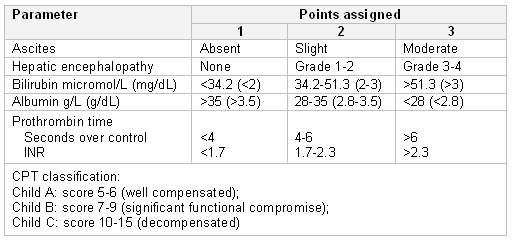
Child-Pugh-Turcotte scoring system
From the collection of Dr Keith Lindor; used with permission
See this image in context in the following section/s:
Videos
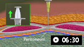 Abdominal paracentesis animated demonstration
Demonstrates how to perform diagnostic and therapeutic abdominal paracentesis.
Use of this content is subject to our disclaimer