Investigations
1st investigations to order
Non-contrast multidetector CT spine (MDCT)
Test
MDCT is the initial imaging modality of choice to evaluate the thoracolumbar spine.[11] Slice thickness should not exceed 5 mm.[11] Thoracolumbar spine imaging may be reconstructed from images concurrently obtained for evaluation of the chest, abdomen and pelvis in a multi-trauma patient. Routine reformatting of these images is not required. A more selective imaging approach is appropriate for patients with a high suspicion of spine injury or when an injury is identified on the non-reformatted images.[11] The sensitivity of CT reaches almost 100%, and it provides details about vertebral fracture morphology (comminution), presence of bony (posterior wall) fragments in the spinal canal, and indirect signs of disruption of the posterior ligamental complex.[10] The main limitation of CT rests in its relative inability to detect changes in soft tissues, including the spinal cord and ligamentous structures.[11][57][58] Despite the high sensitivity of the MDCT in identifying bony abnormalities, interpretation may be difficult in patients with severe degenerative changes or osteopenia.[11]
Result
fractures; increased interpedicular distance, suggesting burst fractures; deformity; subluxation (spondylolisthesis or retrolisthesis)
Investigations to consider
thoracolumbar spine x-ray (anterior-posterior and lateral views)
Test
Although CT has replaced x-rays in most trauma centres, x-rays are usually the first initial diagnostic step.[10] However, spinal fractures frequently go undetected or underestimated on x-rays, especially in the cervical and upper thoracic spine.[10] The European Society for Trauma and Emergency Surgery recommends plain radiographs, guided by the clinical findings, in patients who sustained injuries during low-energy trauma.[10]
Plain radiographs of the cervical and thoracolumbar spine are not recommended by the American College of Surgeons in the initial screening of spinal trauma because of their low sensitivity.[11] CT has a reported sensitivity of 94% to 100% for identifying thoracolumbar spine fractures, whereas radiographs have a reported sensitivity of 49% to 62% for identifying thoracic spine fractures and 67% to 82% sensitivity for identifying lumbar spine fractures.[10][13] Chest and abdominal x-rays taken as part of trauma assessment are not adequate to assess vertebral fractures and alignment.[11][59] If screening of the thoracolumbar spine is performed using radiographs, the American College of Radiology recommends that imaging should consist of anteroposterior and lateral radiographs of the thoracic and lumbar spine with additional 'swimmer’s lateral' view of the upper thoracic spine if this region is obscured by the overlying shoulders.[13]
Clinicians should be alert to the possibility of thoracolumbar fractures if physical abuse of a child is suspected. In physically abused children, 1% of non-accidental head injuries are associated with spinal trauma; 1% to 3% of all fractures in this group of patients are to the vertebrae.[62] One review has recommended that all children with suspected non-accidental injuries who are aged under 2 years should have thoracolumbar x-ray imaging.[63]
Result
fractures; increased interpedicular distance, suggesting burst fractures; deformity; subluxation (spondylolisthesis or retrolisthesis)
MRI spine
Test
Indicated if there are symptoms or signs of spinal cord, conus medullaris, or nerve root injury.[13] MRI is the only modality for evaluating the internal structure of the spinal cord.[11] Useful to identify intramedullary lesions (e.g., post-traumatic cysts, haematomas, oedema) and extramedullary compressions (disc, haematoma, bone fragments). Also useful for assessment of posterior ligamentous complex.[50][66] AOSpine guidelines suggest that when feasible and as long as there are no contraindications, MRI should be used in spinal cord injury patients prior to surgical intervention to improve clinical decision-making, and before or after surgical intervention to predict the neurological outcome.[60] MRI is preferred to CT in children (age <16 years) where cervical injury is suspected.[46][Figure caption and citation for the preceding image starts]: MRI thoracic spine: sagittal view (T2-weighted sequence) showing a pathological fracture of the T10 vertebral body caused by multiple myelomaFrom the personal collection of Dr B. Nurboja and Mr D. Choi [Citation ends].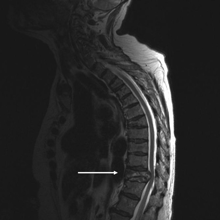 [Figure caption and citation for the preceding image starts]: MRI lumbar spine: sagittal view (T2-weighted sequence) showing an osteoporotic fracture of the T12 vertebral bodyFrom the personal collection of Dr B. Nurboja and Mr D. Choi [Citation ends].
 [Figure caption and citation for the preceding image starts]: MRI lumbar spine: axial view (T2-weighted sequence) showing an osteoporotic fracture of the T12 vertebral bodyFrom the personal collection of Dr B. Nurboja and Mr D. Choi [Citation ends].
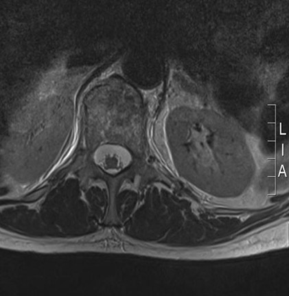
If a new fracture of the spinal column is identified, the rest of the spine should be imaged.[46]
Result
vertebral body fractures, ligamentous complex disruptions, compression of neural elements
CT myelography
Test
Performed when MRI scan of the spine is unavailable or contraindicated (e.g., due to ferromagnetic aneurysm clips, periocular metallic fragments, electronic implants or cardiac pacemakers, haemodynamic instability, severe claustrophobia).
Offers better soft tissue visualisation than normal CT, as well as the ability to visualise disc prolapse.
Result
traumatic disc prolapse; soft tissue damage, haematoma, dural tears
MRI with STIR sequence
Test
Evaluation of patients with compressive vertebral fractures requires performing an MRI including the Short Tau Inversion Recovery (STIR) sequence to distinguish an already healed from an unhealed fracture.
Result
healed or unhealed osteoporotic fracture
Whole body CT
Test
Whole body CT, consisting of scanogram from vertex to toes followed by CT from vertex to mid-thigh, is indicated in adults with multiple injuries with major trauma suspected spinal column injury.[11][46][Figure caption and citation for the preceding image starts]: MRI thoracic spine: sagittal view (T2-weighted sequence) showing a pathological fracture of the T10 vertebral body caused by multiple myelomaFrom the personal collection of Dr B. Nurboja and Mr D. Choi [Citation ends].[Figure caption and citation for the preceding image starts]: MRI lumbar spine: sagittal view (T2-weighted sequence) showing an osteoporotic fracture of the T12 vertebral bodyFrom the personal collection of Dr B. Nurboja and Mr D. Choi [Citation ends].
Result
fractures; soft tissue damage; blunt cerebrovascular injury
Use of this content is subject to our disclaimer