Approach
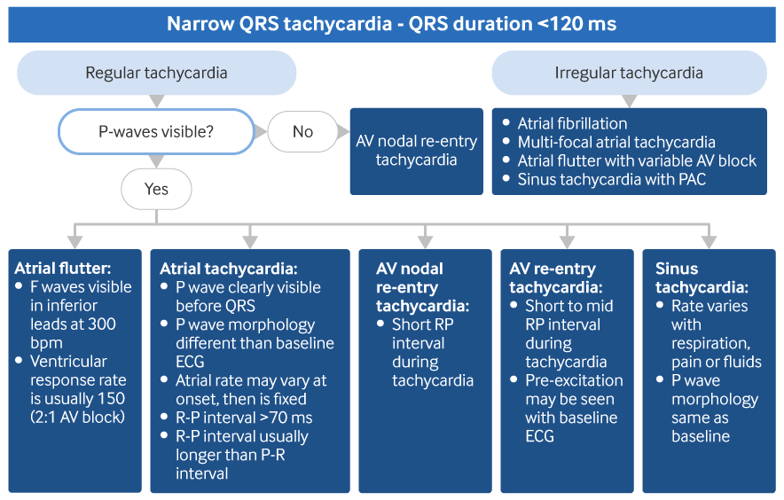
Diagnosis is based on history, the 12-lead ECG, and exclusion of alternative tachyarrhythmic diagnoses through observation of response to manoeuvres. However, inspection of the ECG does not usually reveal the underlying cause.[8][9][Figure caption and citation for the preceding image starts]: Diagnostic algorithm for differentiating atrial tachycardias from other narrow QRS tachycardias. AV = atrioventricular, PAC = premature atrial contractionFrom the collection of Sarah Stahmer, MD [Citation ends].
History/physical
Patients may have a history of prior cardiac disease, COPD, or asthma.[10] Drug history may identify use of digoxin, aminophylline, beta agonists, potassium-wasting diuretics, over-the-counter cold or sinus medications, or substance abuse. A history of recent atrial fibrillation ablation or prior cardiac surgery may be present.[11] There may be symptoms of chest pain, shortness of breath, nausea/vomiting, light-headedness, syncope, palpitations, or fatigue or weakness. Physical examination may detect rales or oedema if congestive heart failure is present.
ECG/Telemetry
The atrial rate in focal atrial tachycardia (focal AT) is typically between 100 and 250 bpm. There is typically a warm-up of the rate for the initial few beats. Often the arrhythmia will occur in short bursts with abrupt onset and cessation.
The ECG should show atrial P waves, and their morphology should be different from the P waves in sinus rhythm. There is typically an isoelectric segment between P waves. At times, irregularity is seen, especially at onset (warm-up) and termination (warm-down).
The PR interval is typically >0.12 seconds (120 milliseconds). The morphology of the P wave in leads aVL and V1 may provide clues about the site of origin. A positive P wave in lead V1 carries 93% sensitivity and 88% specificity for a left atrial focus. In contrast, a positive or biphasic P wave in aVL predicts a right atrial focus with 88% sensitivity and 79% specificity.[12][Figure caption and citation for the preceding image starts]: Focal atrial tachycardia in a 55-year-old with ischaemic cardiomyopathyFrom the collection of Sarah Stahmer, MD [Citation ends].
If the atrial rate is not excessively fast, each P wave will be conducted to the ventricle. If the rate is excessive, or there is AV nodal disease or suppression due to medications, Wenckebach or Mobitz type I second-degree heart block may be seen. In digoxin toxicity, in addition to atrial tachycardia and AV nodal block, there are often signs of abnormal automaticity of non-atrial tissues, such as junctional or ventricular ectopy.
An ambulatory 24-hour (Holter) or longer ECG recording (wearable or implantable loop recorder) may be helpful for diagnosis and management in patients with focal AT that occurs sporadically.[1]
Hypokalaemia alone can predispose non-sinus atrial tissue to spontaneously depolarise, particularly in patients using digoxin.
Vagal manoeuvres or adenosine
An incessant tachycardia that does not show normal variation to pain, respirations, and changes in sympathetic/parasympathetic tone typical of a sinus-based tachycardia indicates a non-sinus origin. Vagal manoeuvres will either have no effect or create an AV nodal block. The nodal block slows the ventricular response rate but has no effect on the atrial rate. Adenosine causes AV node blockade and may transiently suppress, or more rarely terminate, the tachycardia.[13][14]
Vagal manoeuvres or adenosine may help differentiate atrial tachycardia from other causes of supraventricular tachycardia, however this information can be misleading or limited if the tachycardia terminates.[Figure caption and citation for the preceding image starts]: Response to adenosine 6 mg intravenouslyFrom the collection of Sarah Stahmer, MD [Citation ends].
The differential diagnosis of atrial tachycardia will often include sinus tachycardia, AV node re-entrant tachycardia, AV re-entrant tachycardia or atrial flutter.
Sinus tachycardia will transiently slow in response, with slowing of the sinus rate and prolongation of the PR interval before AV block (if it occurs).
AV node re-entrant tachycardia or AV re-entrant tachycardia (accessory pathway mediated supraventricular tachycardia) will abruptly cease, with resumption of sinus rhythm after a pause.
In atrial flutter there will be transient slowing of the ventricular response rate with AV block. The characteristic flutter waves will be revealed and will be unaffected.
This is not a foolproof therapeutic intervention, as some atrial tachycardias, particularly those due to micro re-entry, will break in response to these manoeuvres. Most will show no response or show transient AV block with persistent atrial depolarisation (P waves).
Further tests
Serum analysis should follow clinical suspicion of aetiology. Electrolytes should be routine. Levels of medications that can predispose patients to these rhythms can be ordered when appropriate, specifically theophylline and digoxin. When drugs of abuse are suspected, specifically cocaine, amphetamines, and ethanol, quantitative supportive data can be valuable. Thyroid-stimulating hormone (TSH) level helps rule out suspected hyperthyroidism.
Echocardiography can be helpful in patients for whom the underlying cause is suspected to be due to previously undiagnosed cardiac pathology, such as valvular disease and cardiomyopathies. Patients with evidence of haemodynamic compromise or heart failure should have an echo performed as soon as possible.
Electrophysiological study (EPS) is an invasive test that can assist in confirming the origin and site of arrhythmogenesis for selected patients for whom the diagnosis is unclear or who are not responding to empirical therapies. It can be curative if coupled with ablation.[1]
Use of this content is subject to our disclaimer