Etiology
MCL injuries require that a valgus (twisting outward away from midline) and/or external rotation load be placed on the knee. Valgus loads can occur through contact, noncontact, and overuse mechanisms.[9] Contact mechanisms, commonly associated with a blow to the lateral knee, involve large valgus stresses and often result in a complete MCL tear. Noncontact valgus and external rotational stresses, observed in cutting, pivoting, and deceleration motions of the leg (e.g., skiing), frequently cause partial tears of the MCL. Finally, overuse mechanisms may be observed in sports such as swimming (breaststroke whip kick) and gymnastics, which place repetitive valgus loads across the knee joint.[13]
Pathophysiology
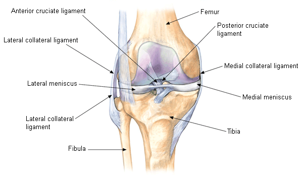
Anatomically, the MCL is composed of a superficial and a deep portion. The superficial MCL resides in the middle layer of the medial compartment of the knee and spans from the medial femoral condyle to its broad insertion at the metaphysis of the tibia, 4 to 5 cm below the joint line. [Figure caption and citation for the preceding image starts]: Anterior knee anatomy (right knee), patella removedCreated by Sanjeev Bhatia, MD; used with permission [Citation ends]. [Figure caption and citation for the preceding image starts]: Oblique view of medial knee (right knee). MCL: medial collateral ligamentCreated by Sanjeev Bhatia, MD; used with permission [Citation ends].
 It is the principal restraint to valgus forces at the knee joint at all degrees of flexion. The deep MCL resides in the third layer of the medial compartment and is often separated from the superficial MCL by a bursa, which facilitates sliding of the two MCL components during flexion. The deep MCL attaches to the medial meniscus but does not aid in resisting valgus stress on the knee.
The severity of MCL injury is proportionally related to the number of ligament fibers torn in the superficial MCL. The most common location for MCL injuries is the femoral insertion.[13] Grade I injuries consist of a minimal number of torn fibers, grade II injuries have an increased degree of ligamentous disruption, and grade III injuries result in complete tearing of MCL fibers. Paradoxically, higher-grade MCL injuries are usually associated with less pain, perhaps because there is little or no tension on the injured ligament.[13] Patients with MCL injuries repeatedly experience a sense of valgus instability and altered biomechanics leading to post-traumatic arthrosis. Healing of grade I and II MCL injuries usually follows a relatively predictable sequence: hemorrhage, inflammation, proliferation, and remodeling.[14]
Patients frequently have concomitant injury to the anterior cruciate ligament, posterior cruciate ligament, and meniscus. See Anterior cruciate ligament (Diagnostic approach) and Meniscal tear (Diagnostic approach).
In knee injuries from cutting maneuvers (occurring when an athlete forcefully changes direction on a weight-bearing foot), valgus forces placed on the knee usually tear the medial capsular ligament first, then the MCL, and finally the anterior cruciate ligament.[15] Because the meniscus is firmly attached to the deep MCL, meniscal tears commonly present alongside MCL tears.
Classification
O'Donoghue classification[1]
Isolated grade I MCL injury (mild)
MCL has a few torn fibers but there is no loss of ligamentous integrity.
Isolated grade II MCL injury (moderate)
MCL is partially torn. However, the fibers are still opposed. There might be mild pathologic laxity, which may or may not be symptomatic.
Isolated grade III MCL injury (severe)
Integrity of the MCL is completely disrupted. There is significant pathologic laxity of the knee with valgus stress.
American Medical Association Committee on the Medical Aspects of Sports standard nomenclature of athletic injuries[2]
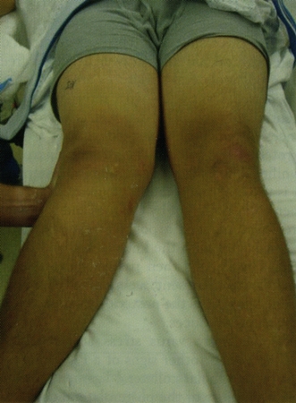
MCL injuries are classified based on the amount of medial joint opening when a valgus load is applied at 20 to 30 degrees of knee flexion: [Figure caption and citation for the preceding image starts]: The abduction (valgus) stress testFrom the collection of Sanjeev Bhatia, MD; used with permission [Citation ends].
Grade I: 0 to 5 mm of opening
Grade II: 5 to 10 mm of opening
Grade III: >10 mm of opening
Proposed classification, based on MRI findings:[3]
Type 1: Pre-avulsion injury
Type 2: Avulsion injury
Type 3: MCL injury
Type 4: Distal rupture of the MCL and bone contusion
Other forms of MCL injury
MCL + anterior cruciate ligament (ACL) injury
The ACL is the most commonly injured ligament along with the MCL.
MCL + multiligament injury
The posterior cruciate ligament, lateral collateral ligament, and menisci are frequently simultaneously injured.
Recurring MCL injury
Recurring, or chronic, MCL injury is a complication of acute MCL injury.
Use of this content is subject to our disclaimer