Approach
A meniscal tear is the most common soft tissue injury affecting the knee. The meniscus is the soft fibrous shock absorber in the knee. The lateral meniscus covers 84% of the lateral tibial plateau; the medial meniscus covers 64% of the medial tibial plateau.[23]
A meniscus is at risk of tearing when experiencing abnormal forces, usually during sporting activities, and bending and twisting motions. Loss of the meniscus diminishes the area of force distribution, increases force concentration, and eventually leads to arthritis. Torn menisci rarely heal spontaneously. Early diagnosis can lead to effective surgical repair and joint preservation.
Meniscal tears frequently occur in conjunction with knee ligament injuries, especially of the anterior cruciate ligament (ACL). The Scandinavian ACL registries showed that 35% to 55% of ACL injuries are combined with meniscal injury.[24][Figure caption and citation for the preceding image starts]: Types of meniscal tearsCreated by BMJ Publishing Group [Citation ends].
History
Mechanism of injury, location of pain, associated knee symptoms, and any previous knee injuries (especially ACL injury) are important details to elicit on history taking. Patients typically report twisting of the affected knee. Knee swelling, usually occurring several hours after the injury, is common but not always present. Patients often describe a sensation of the femur and tibia twisting apart from each other (catching or locking) or the knee feeling loose, indicating knee instability or buckling. Knee pain varies greatly; some may experience minimal pain, while others describe only intermittent pain.
It is also important to establish the presence of risk factors, including level of patient activity (sporting and non-sporting), history of knee joint arthritis, or any other knee conditions (e.g., malalignment of the knee joint or known knee instability). Details regarding playing surfaces and weather conditions should also be discussed, if applicable.
Physical examination
On inspection, the knee joint may or may not be swollen. Swelling at the posterior aspect of the knee suggests a popliteal (Baker's) cyst. Popliteal cysts are associated with meniscal pathology in 80% of cases.[25] See Popliteal cyst for more information. Joint line crepitation and tenderness directly over the joint line is a common finding on palpation. Range of motion may or may not be limited.
The following clinical tests may be performed specifically to evaluate for a meniscal tear.[1][2]
Palpation of the joint line: the patient is in the supine position with the knee in flexion. The examiner pushes the joint line between femur and tibia. Pain by the palpation suggests a meniscal tear.
Bounce home test: the examiner moves the knee from a flexed position to an extended position. Pain in the extended position suggests a meniscal tear. This test is sensitive but not specific for a torn meniscus. Palpation and bounce home test Opens in new window
McMurray's test: the patient is in the supine position with the knee in flexion. The examiner flexes the hip and, with one hand on the joint line, rotates the foot internally and externally. Pain with rotation suggests a meniscal tear. This test has low sensitivity and high specificity for diagnosing a meniscal tear. McMurray test Opens in new window[26]
Ege's test: also known as the weight-bearing McMurray's test. The patient is asked to slowly squat with both knees in maximal external rotation. Discomfort or clicking in the knee suggests a positive test. Not suitable for patients with a traumatic tear due to the need to weight bear.
Apley's test: performed with the patient prone and the knee flexed at 90°. The examiner places an axial load on the lower leg while rotating the foot. The patient often feels pain in the affected compartment. Apley test Opens in new window[27]
Hyperextension test: the examiner lifts the heel of the affected leg, hyperextending the knee and adding additional downwards force on the tibia. The patient reports pain in the affected compartment. This test is sensitive but not specific for a torn meniscus.
Thessaly's test: the patient stands on one leg while the examiner flexes the knee to 5° and rotates medially and laterally three times. The test is then repeated with the knee flexed at 20°. Discomfort or a sense of locking or catching in the knee suggests a positive test.
Investigations
A magnetic resonance imaging (MRI) scan is considered the ideal imaging test for meniscal tear.[3][28] Findings are highly sensitive and specific, documenting increased signal within the substance of the meniscus and demonstrating conformational changes of the meniscus cartilage. In addition, an MRI scan may be useful for identifying associated ligament compromise and articular cartilage changes, as well as supporting pre-surgical planning if arthroscopy is indicated.[1]
X-ray is indicated in patients with a history of arthritis and chronic long-standing meniscal tears where Fairbanks' changes may be found, such as flattening of the femoral condyle, narrowing of the joint space, and osteophytic ridging.[3] Recommended x-ray series include standing anterior-posterior x-ray, lateral knee x-ray, 45° posteroanterior flexion, and skyline views. If the leg is malaligned, then a long-leg hip to ankle film should also be obtained.
Arthroscopy, although usually performed to repair or resect the torn meniscus, can be used to confirm the diagnosis. [Figure caption and citation for the preceding image starts]: MRI scan demonstrating horizontal cleavage tear of medial meniscus (arrow); white horizontal line separates the inferior and superior portions of the torn meniscusFrom the collection of Dr Kevin R. Stone [Citation ends].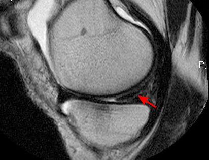 [Figure caption and citation for the preceding image starts]: Arthroscopic view of horizontal cleavage tear of lateral meniscus (arrow)From the collection of Dr Kevin R. Stone [Citation ends].
Use of this content is subject to our disclaimer