History and exam
Key diagnostic factors
common
injury due to excessive or repetitive valgus loading of MCL
Lower-grade MCL injuries frequently occur with noncontact valgus and external rotation injuries (e.g., twisting injuries during skiing accidents).
More severe injuries are usually associated with a blow to the lateral side of the knee.
medial knee pain
MCL injury is associated with pain on the medial side of the knee along the length of the ligament, both above and below the joint line.
Paradoxically, higher-grade MCL injuries are usually associated with less pain, perhaps because there is little or no tension on the injured ligament.[15]
joint effusion
The location of joint effusion has been shown to correlate with the injury site in the superficial MCL 64% of the time.[15]
Absence of joint effusion may indicate a severe tear: grade III injuries result in tearing of the joint capsule, allowing fluid to escape to surrounding soft tissues.
Speed of onset of swelling provides clues to the pathology involved. An acute effusion, within 2 hours of injury, suggests hemarthrosis. Swelling 12 to 24 hours after injury usually indicates a synovial effusion.[18]
Hemarthrosis, while uncommon, is suspicious for injury to the anterior cruciate ligament.
tenderness
Usually occurs at the adductor tubercle or proximal tibia. In 76% of cases, the site of tenderness corresponds with the site of injury in the superficial MCL.[15] Medial meniscal tears have tenderness limited to the joint line.
laxity on valgus stress testing
The abduction stress test (i.e., applying a valgus load to the knee) at 30 degrees flexion is an excellent diagnostic tool. [Figure caption and citation for the preceding image starts]: The abduction (valgus) stress testFrom the collection of Sanjeev Bhatia, MD; used with permission [Citation ends].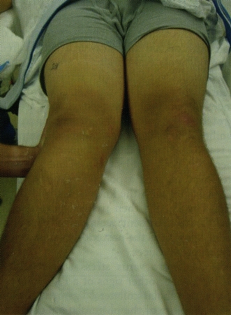 Pain and disproportionate laxity imply stretching or tearing of the MCL.
Pain and laxity with valgus stress in a fully extended knee suggest coexistent anterior cruciate ligament tear.[18]
Other diagnostic factors
common
ecchymosis
Ecchymosis over the MCL often develops 1 to 3 days after injury.
uncommon
audible pop or tearing sensation at time of injury
Suggests grade III MCL injury or cruciate ligament injury.
difficulty walking
Most patients are able to continue walking after an acute injury. In one study, even with grade III MCL injuries, 76% of patients were able to walk into an office unaided by external support.[15]
instability symptoms of knee
Most MCL injuries are not associated with instability symptoms ("giving way"). The rare exception is injury to the posterior oblique portion of the deep MCL, which can result in anteromedial instability.
If instability is a prominent feature, anterior cruciate ligament or posterior cruciate ligament injury is likely.
mechanical knee symptoms
Symptoms such as catching, locking, giving way, and popping are not usually associated with MCL injuries. Concomitant meniscal tear or cruciate ligament injury should be suspected.
knee deformity
May signify patellar subluxation or dislocation.
positive anterior drawer test
Test is performed with the foot in external rotation and knee flexed at 90 degrees.
Any anteromedial instability evident during test [Figure caption and citation for the preceding image starts]: The anterior drawer testFrom the collection of Sanjeev Bhatia, MD; used with permission [Citation ends].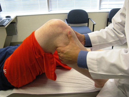 suggests that the deep MCL may be damaged, specifically the posterior oblique portion.
positive posterior drawer test
Test is performed the same way as an anterior drawer test but with a push on the tibia.
Any laxity indicates injury to the posterior cruciate ligament.
positive Lachman test
The best diagnostic test for anterior cruciate ligament (ACL) injury. [Figure caption and citation for the preceding image starts]: The Lachman testFrom the collection of Sanjeev Bhatia, MD; used with permission [Citation ends].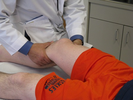
The patient's knee is put in 20 to 30 degrees of flexion. One hand is placed on the patient's thigh and the other behind the tibia (with the thumb on the tibial tuberosity). The tibia is pulled anteriorly.
The incidence of ACL tears has been found to be 20% when there is no valgus laxity on clinical exam, 53% with valgus laxity in 30 degrees of flexion, and 78% with valgus laxity in full knee extension.[5]
positive pivot shift test
Used in conjunction with the Lachman test for diagnosing anterior cruciate ligament (ACL) injury.
An internal rotation and valgus stress is applied to the knee while taking it from 20 to 40 degrees of flexion. In an ACL-deficient knee, the tibia will anterolaterally sublux in the initial phase of flexion and then reduce with further flexion.
joint line tenderness
Although not the most sensitive or specific sign for meniscal injury,[26] joint line tenderness may indicate potential meniscus pathology.
chronic pain
Knowing when the patient injured his or her knee helps distinguish acute from chronic MCL injury. A chronic MCL injury is one that has occurred ≥3 months previously.[4]
Risk factors
strong
participation in activities involving valgus stress at the knee joint
Valgus loads - occurring through contact, noncontact, or overuse mechanisms - are required for straining the MCL. Typically, a valgus load can be in the form of a lateral blow to the lower thigh or external rotation of the tibia relative to the femur. People who frequently experience these stresses on the knee (e.g., participants in American football, rugby, hockey, skiing) are most at risk.[5][16][17][18]
age 20 to 35 years
age 55 to 70 years
Older, less active adults are prone to MCL injury during falls.[18]
weak
weak muscles that cross the medial aspect of knee
Having weak muscles in the posterior aspect of the knee - pes anserinus mainly, but also the semimembranosus - may decrease the dynamic stability of the knee joint.[18] There is no firm evidence that this increases the likelihood of MCL injury.
Use of this content is subject to our disclaimer