Tests
1st tests to order
plain x-rays of knee
Test
Ordered in accordance with the Ottawa knee rules to exclude bony injury. [Figure caption and citation for the preceding image starts]: X-ray indications in acute knee injury: the Ottawa knee rulesTable created by Sanjeev Bhatia, MD. Adapted from Stiell IG, et al. Implementation of the Ottawa knee rule for the use of radiography in acute knee injuries. JAMA. 1997;278:2075-2079 [Citation ends].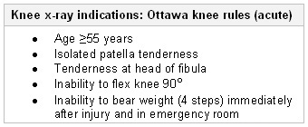 [27] Anteroposterior, lateral, and patellofemoral views are often sufficient.
Pellegrini-Stieda lesions (a calcification that develops adjacent to the adductor tubercle) suggest a collateral ligament injury that is more than 6 weeks old. They are best seen in the anteroposterior view.
Result
may show associated fracture of the tibial plateau, patella, or distal femur; calcification adjacent to the adductor tubercle is typical of a Pellegrini-Stieda lesion in chronic situations
stress x-rays of knee
Test
Stress radiography x-rays taken with a valgus load on the knee at 20 degrees of flexion should be ordered in adolescents to rule out physeal injuries and also in adults to objectively define the amount of medial compartment gapping.
Result
greater than normal opening on the medial side of the knee joint is commonly seen; physeal fractures may be seen in adolescents; in adults, a greater than 3.2 mm side-to-side difference of medial gapping is suggestive of a grade III MCL injury
Tests to consider
MRI of knee
Test
Provides excellent visualization of soft tissue anatomy and is indicated if any associated injuries are suspected.[27]
MRI is also useful for identifying the precise location of the MCL tear, which is usually visible on T2-weighted images.[Figure caption and citation for the preceding image starts]: T2-weighted MRI showing a medial collateral ligament injuryFrom the collection of Sanjeev Bhatia, MD; used with permission [Citation ends].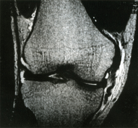
Result
with an MCL tear, high signal edema and hemorrhage may be seen in the low-signal ligament; may also show meniscal tear, anterior cruciate ligament or posterior cruciate ligament tear, bone bruise, osteochondral fracture
Emerging tests
diagnostic ultrasound
Test
Ultrasound is an excellent and efficient means for visualizing the knee.[27] MCL tears, as well as associated injuries, may be visualized with a knee ultrasound. However, grade III MCL injuries (complete tears) are difficult to diagnose with ultrasound because of the irregular nature of ligament tearing.[24]
Result
MCL appears thickened and hypoechoic (from edema); fluid collection may be greatest near the site of the tear; Pellegrini-Stieda lesions appear as calcifications within thickened and hypoechoic ligament tissue
Use of this content is subject to our disclaimer