Differentials
Oral squamous cell carcinoma
SIGNS / SYMPTOMS
May be associated lymphadenopathy or dysphagia.
Pain or numbness indicates deep invasion into bony structures or soft tissues.
INVESTIGATIONS
Incisional biopsy and pathology demonstrates evidence of invasive carcinoma; keratin pearls may be seen.
Chronic candidiasis
SIGNS / SYMPTOMS
The tissue response to chronic candidal infection is hyperplastic in nature - in particular, in the retrocommissural area, where a more speckled to atrophic surface pattern is often present.
In the immunosuppressed patient and in cases of T-cell dysfunction this finding assumes greater clinical importance, as it concerns potential malignant transformation within the speckled areas. [Figure caption and citation for the preceding image starts]: Candidal leukoplakiaCourtesy of Dr James Sciubba; used with permission [Citation ends].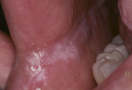
INVESTIGATIONS
Candidal smear is positive.
Incisional biopsy and pathology may demonstrate a range of features, from benign hyperplasia with hyperkeratosis in most circumstances, to variable degrees of dysplasia or invasive cancer less frequently. Surface Candida hyphae may be seen.
Submucous fibrosis
SIGNS / SYMPTOMS
Underlying tissues are firm and inelastic.
INVESTIGATIONS
Incisional biopsy and pathology will demonstrate epithelial atrophy and fibrosis of the underlying lamina propria.
Hairy leukoplakia
SIGNS / SYMPTOMS
Painless white plaques along the lateral tongue borders.
History of HIV or immunosuppression.[Figure caption and citation for the preceding image starts]: Oral hairy leukoplakiaCourtesy of Dr James Sciubba; used with permission [Citation ends].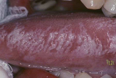
INVESTIGATIONS
Definitive diagnosis is through biopsy and histologic evaluation of the lesion.
In situ hybridization technique demonstrates the presence of Epstein-Barr virus in the tissue.
Syphilitic leukoplakia
SIGNS / SYMPTOMS
Typically located predominantly over the dorsum of the tongue, in contrast to the lateral and ventral surfaces, where the vast majority of oral leukoplakia are found; plaques of keratinized tissue with fissured topography are characteristic.[132]
INVESTIGATIONS
Treponemal-specific serology tests are antigen-based tests and remain positive lifelong if current or past infection: tests include treponemal enzyme immunoassay, Treponema pallidum particle agglutination, T pallidum hemagglutination, fluorescent antibody absorption tests, and immunocapture assay.
Nontreponemal titers Venereal Disease Research Laboratory or rapid plasma reagin correlate with disease activity, decreasing or becoming nonreactive with effective treatment.
Frictional keratosis
SIGNS / SYMPTOMS
May be able to identify a source of chronic irritation (e.g., a faulty dental restoration, an ill-fitting denture, or parafunctional habits such as bruxism or chronic cheek biting).
Removal of offending trauma may lead to resolution of keratosis.
INVESTIGATIONS
Definitive diagnosis is through biopsy and histologic evaluation of the lesion.
Lichen planus
SIGNS / SYMPTOMS
Typically presents with reticular, atrophic, and erosive mucosal changes in a symmetrical distribution. Reticular/plaque lesions are usually asymptomatic; erosive lesions may be painful.
Lichenoid lesions elsewhere on the skin may be present.
INVESTIGATIONS
Incisional biopsy and pathology may demonstrate characteristic superficial keratinization, dense banded lymphocytic infiltrate within the superficial lamina propria, and liquefactive basal layer degeneration and scattered colloid bodies or apoptotic keratinocytes.
Epithelial dysplasia may be a feature. Malignant transformation is well recognized; the atrophic and erosive forms are reported to have the highest rate of malignant transformation, with published rates of malignant transformation of oral lichen planus ranging from 0% to 2%.[133]
Discoid lupus erythematosus
SIGNS / SYMPTOMS
Typically presents with a lichenoid pattern of keratosis and erosive/inflammatory lesions. Most often the lesions are accompanied by radiating reticular striaform lesions; plaque-like lesions may also be seen but are less common.[134]
However, in some cases, oral lesions with plaque-like configuration similar to leukoplakia may be evident in spite of quiescent disease elsewhere.[135]
INVESTIGATIONS
ANA and antidouble-stranded DNA antibody positive.
Incisional biopsy and pathology may demonstrate vacuolated keratinocytes, patchy periodic acid-Schiff-positive patches and edema in the lamina propria, and severe or perivascular inflammatory infiltration.[136] Direct immunofluorescence will show an uneven globular deposition of IgG, IgA, and fibrinogen along the basement membrane zone.[137]
White sponge nevus
SIGNS / SYMPTOMS
Rare inherited (autosomal dominant) developmental abnormality characterized by white plaques present on the buccal mucosa (often bilateral), and less commonly the lingual and labial tissues; vagina, rectum, and nasal cavity may also be affected. Often detected during childhood.
INVESTIGATIONS
No differentiating test is usually performed as clinical findings alone are usually adequate.
Use of this content is subject to our disclaimer