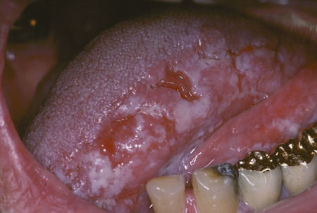
Oral leukoplakia presents as white plaques of questionable risk, diagnosed when other known diseases or disorders that carry no risk for oral cancer have been excluded.
Multiple clinical forms exist: homogeneous, speckled, nodular, and verrucous.
May be idiopathic, but is commonly seen in heavy tobacco users and consumers of alcohol or areca nut (betel quid).
The majority are histologically benign with a wide range of histologic characteristics. Grading of dysplasia assists in selecting management options.
Certain leukoplakias, particularly nonhomogeneous leukoplakias, such as speckled leukoplakia and proliferative verrucous leukoplakia, have a significant risk of malignant transformation. They require habit intervention and frequent and careful follow-up, often with biopsy confirmation or definition of the biologic nature of the leukoplakia over time.
Oral leukoplakia, as traditionally defined by the World Health Organization (WHO), is a predominantly white lesion of the oral mucosa that cannot be characterized as any other definable lesion.[1]Kramer IR, Lucas RB, Pindborg JJ, et al. Definition of leukoplakia and related lesions: an aid to studies on oral precancer. Oral Surg Oral Med Oral Pathol. 1978 Oct;46(4):518-39.
http://www.ncbi.nlm.nih.gov/pubmed/280847?tool=bestpractice.com
[2]Axell T, Holmstrup P, Kramer IRH, et al. International seminar on oral leukoplakia and associated lesions related to tobacco habits. Community Dent Oral Epidemiol. 1984 Jun;12(3):145-54.[3]Axell T, Pindborg JJ, Smith CJ, et al. Oral white lesions with special reference to precancerous and tobacco-related lesions: conclusions of an international symposium held in Uppsala, Sweden, May 18-21 1994. International Collaborative Group on Oral White Lesions. J Oral Pathol Med. 1996 Feb;25(2):49-54.
http://www.ncbi.nlm.nih.gov/pubmed/8667255?tool=bestpractice.com
Leukoplakia is often associated with tobacco smoking, although idiopathic forms are not rare.[2]Axell T, Holmstrup P, Kramer IRH, et al. International seminar on oral leukoplakia and associated lesions related to tobacco habits. Community Dent Oral Epidemiol. 1984 Jun;12(3):145-54.[3]Axell T, Pindborg JJ, Smith CJ, et al. Oral white lesions with special reference to precancerous and tobacco-related lesions: conclusions of an international symposium held in Uppsala, Sweden, May 18-21 1994. International Collaborative Group on Oral White Lesions. J Oral Pathol Med. 1996 Feb;25(2):49-54.
http://www.ncbi.nlm.nih.gov/pubmed/8667255?tool=bestpractice.com
An international working group has amended the earlier WHO definition as follows: "The term leukoplakia should be used to recognize white plaques of questionable risk having excluded (other) known diseases or disorders that carry no risk for cancer".[4]Warnakulasuriya S, Johnson NW, van der Waal I. Nomenclature and classification of potentially malignant disorders of the oral mucosa. J Oral Pathol Med. 2007 Nov;36(10):575-80.
http://www.ncbi.nlm.nih.gov/pubmed/17944749?tool=bestpractice.com
[5]Warnakulasuriya S, Kujan O, Aguirre-Urizar JM, et al. Oral potentially malignant disorders: a consensus report from an international seminar on nomenclature and classification, convened by the WHO Collaborating Centre for Oral Cancer. Oral Dis. 2021 Nov;27(8):1862-80.
http://www.ncbi.nlm.nih.gov/pubmed/33128420?tool=bestpractice.com
Leukoplakias are commonly homogeneous and most are benign. Nonhomogeneous leukoplakia, or so-called erythroleukoplakias, for example speckled leukoplakia or nodular leukoplakia - predominantly white or white and red lesions with an irregular texture that may be flat, nodular, exophytic, or papillary/verrucous - are more likely to be potentially malignant. Histologic features of both forms of leukoplakia are variable and may include orthokeratosis or parakeratosis of various degrees, mild inflammation, and variable degrees of epithelial dysplasia. However, although criteria for dysplasia have been defined by the WHO, it is difficult to make an objective categorization of dysplasia owing to a high inter-observer and intra-observer variation in assessment.[6]El-Naggar AK, Chan JKC, Grandis JR, et al, eds. WHO classification of head and neck tumours. 4th ed. Lyon, France: International Agency for Research on Cancer; 2017.[7]Speight PM, Khurram SA, Kujan O. Oral potentially malignant disorders: risk of progression to malignancy. Oral Surg Oral Med Oral Pathol Oral Radiol. 2018 Jun;125(6):612-27.
https://www.oooojournal.net/article/S2212-4403(17)31248-8/fulltext
http://www.ncbi.nlm.nih.gov/pubmed/29396319?tool=bestpractice.com