Etiology
The etiology of oral leukoplakia is multifactorial and some are idiopathic. The most commonly associated risk factor is the use of tobacco in either smoked or smokeless forms. Additionally, the use of areca nut (betel quid) preparations in many parts of the world (generally South and Southeast Asia) poses a significant risk, as does the use of dry snuff and other forms of smokeless tobacco. There is weak evidence on chronic (excess) alcohol use as a risk factor for oral leukoplakia. The role of chronic candidiasis has been linked to the development of leukoplakia: in particular, nonhomogeneous leukoplakia. This is possibly related to the high nitrosation potential of some candidal forms, suggesting endogenous nitrosamine production.[10] The habit of reverse smoking (lit end retained within the mouth) in some cultures can produce a wide range of oral mucosal lesions, including leukoplakia of the palate. In these populations such leukoplakias have a 19-times increased risk of malignant transformation compared with those where tobacco is used in other forms.[11] Sanguinaria-associated white lesions or keratoses have also been described, which may be extensive and multifocal; these may resemble and be misinterpreted as proliferative verrucous leukoplakia.[12] In cases of proliferative verrucous leukoplakia, a multifocal and often recalcitrant form of leukoplakia, the usual risk factors regarding etiology and risk of malignant transformation, including tobacco habits and alcohol consumption, do not apply.[13][14] High-frequency allelic loss and high-risk allelic profiles have been noted in an attempt to account for high-risk progression to dysplasia and malignant transformation.[15]
Pathophysiology
The primary pathologic change present within leukoplakia is that of abnormal epithelial differentiation and growth proliferation with increased surface keratinization producing the clinical appearance of a whitened mucosa. This may be accompanied by alterations in the epithelial thickness. For example, the epithelium may show atrophy or acanthosis (thickened prickle cell layer), and both features may exist within a single lesion. In erythroleukoplakia, the clinical appearance of a mixed red-and-white patch is a result of epithelial atrophy and a reduction/absence of surface keratin.
From a clinical management perspective, however, the most significant changes are those of epithelial dysplasia that may exist as a component of the altered epithelial proliferation and differentiation.
The underlying molecular basis for such changes is uncertain.[16] However, some data exist from research in premalignant forms of leukoplakia centered on genetic alterations in oncogene/tumor suppressor gene expression that may herald dysplastic transformation or serve as diagnostic or therapeutic targets.[17] Other factors include loss of differentiation-related keratins and carbohydrate antigens.[18] Oxidative stress and nitrative DNA damage via reactive nitrogen products such as inducible nitric oxide synthase and inflammation-related mechanisms have also been implicated in oral leukoplakia and its transformation to dysplasia and carcinoma.[19] Molecular marker studies have demonstrated that leukoplakia lesions have an increase in cells that stain positively for p53 and the proliferation marker Ki67.[20] Several studies have shown other genetic changes including alterations in keratin gene expression, cell cycle alteration, and increases in loss of heterozygosity aneuploidy within oral leukoplakia.[18][21][22][23] However, the most critical and useful marker to guide clinical management is the confirmed presence and grading of epithelial dysplasia due to its relationship to carcinomatous transformation.[24]
Classification
Clinical classification
Three main types of oral leukoplakia are described clinically:
1. Homogeneous oral leukoplakia [Figure caption and citation for the preceding image starts]: Homogeneous leukoplakiaCourtesy of Dr James Sciubba; used with permission [Citation ends].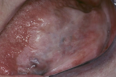
Most common type
Presents as a uniformly white plaque
Occurs mainly on buccal mucosa or lateral margin of tongue
Low transformation potential.
2. Nonhomogeneous leukoplakia
Speckled leukoplakia [Figure caption and citation for the preceding image starts]: Speckled leukoplakiaCourtesy of Dr James Sciubba; used with permission [Citation ends].
Less common
More serious than the homogeneous form
Consists of white flecks or fine nodules on an atrophic erythematous base
Stronger malignant potential than homogeneous leukoplakia
Regarded as a combination of or a transition between leukoplakia and erythroplasia; hence, the new term erythroleukoplakia in the new World Health Organization classification.[5]
Nodular leukoplakia
Small aggregated hemispherical red or white surface alterations or excrescences
May show a red background or substrate
Stronger risk of dysplasia or malignant potential than in homogeneous leukoplakia.
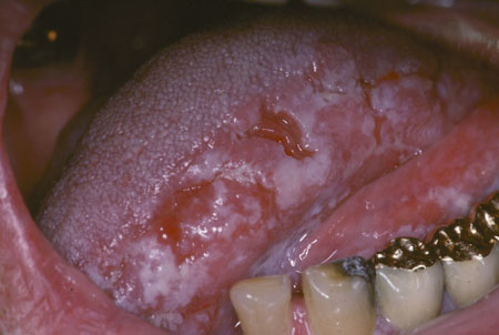
3. Proliferative verrucous leukoplakia [Figure caption and citation for the preceding image starts]: Proliferative verrucous leukoplakiaCourtesy of Dr James Sciubba; used with permission [Citation ends].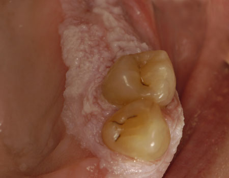
Least common type of oral leukoplakia
High risk of intervening dysplasia and carcinoma developing
Progressive, widespread, and multifocal in nature
Often affects the gingiva
High rate of recurrence following excision and histologic progression toward carcinoma.
Causes of nonleukoplakia oral white lesions
Local causes:
Materia alba (debris from poor oral hygiene)
Keratoses (e.g., frictional keratosis [and cheek/lip biting], smoker's keratosis of palate [leukokeratosis nicotina palati])
Chemical burns
Skin grafts
Scars
Furred or hairy tongue
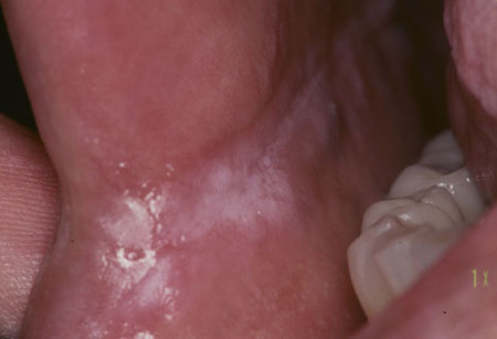
Developmental/inherited disorders of keratinization (e.g., white sponge nevus). [Figure caption and citation for the preceding image starts]: Snuff-associated leukoplakia (snuff "pouch")Courtesy of Dr James Sciubba; used with permission [Citation ends].
Neoplasms:
Carcinoma
Verruciform xanthoma.
Infections:
Candidiasis (thrush: candidal leukoplakia)
Epstein-Barr virus (hairy leukoplakia)
Syphilis (syphilitic mucous patches and keratosis)
Measles (Koplik spots).[Figure caption and citation for the preceding image starts]: Candidal leukoplakiaCourtesy of Dr James Sciubba; used with permission [Citation ends].
 [Figure caption and citation for the preceding image starts]: Oral hairy leukoplakiaCourtesy of Dr James Sciubba; used with permission [Citation ends].
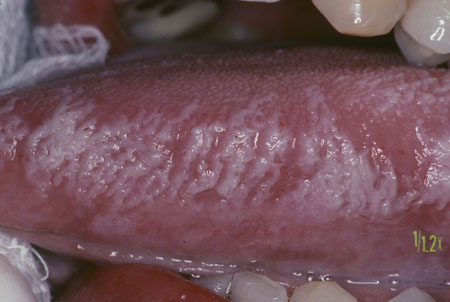
Mucocutaneous disease:
Lichen planus
Lupus erythematosus.
Use of this content is subject to our disclaimer