Tests
1st tests to order
chest x-ray
Test
Volumes of less than 50 mL of pleural gas may be visible in the upright position. At least 100 mL of intrapleural gas is necessary to visualize a pneumothorax in supine patients.[46][Figure caption and citation for the preceding image starts]: Anterior-posterior chest x-ray demonstrating a right pneumothoraxFrom the collection of Dr Ryland P. Byrd [Citation ends].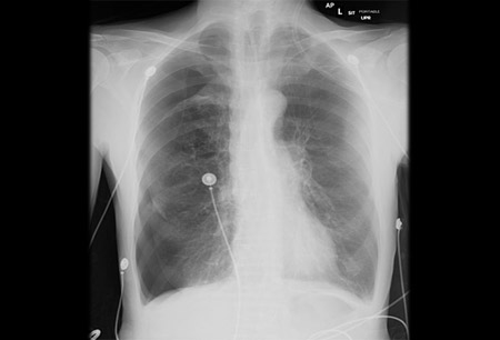
Presence of visceral pleural line, lung atelectasis, and loss of volume may suggest pneumothorax ex vacuo.[45]
Result
visceral pleural line typically identified; if the patient has an underlying lung disease, other abnormalities such as emphysema, lung masses, and pulmonary infiltrates may be present
chest ultrasound
Test
In patients with trauma or blunt chest injuries, chest ultrasound has become the initial test of choice due to rapid results and overall superior performance to a single view chest x-ray (CXR).[42] In one Cochrane review, chest ultrasound had a sensitivity of 91% and a specificity of 99% (compared with a sensitivity of 47% and a specificity of 100% for CXR) for detecting pneumothoraces in trauma patients in the emergency department. The authors concluded that chest ultrasound is superior to supine CXR, independent of the type of trauma, type of chest ultrasonography operator, or type of chest ultrasonography probe used, and they recommended that chest ultrasound is included in trauma protocols (e.g., advanced trauma life support [ATLS]).[43]
Result
absent lung sliding
Tests to consider
CT chest
Test
A CT scan of the chest is more sensitive than a chest radiograph or ultrasound in detecting pneumothoraces. It is often used in patients with multiple traumatic injuries or where an occult pneumothorax is suspected.[44]
CT scanning may also be necessary in patients with underlying respiratory diseases. CT is very useful in differentiating a pneumothorax from bullous emphysema.[41][45]
Result
visceral pleural line easily identified; possibly atelectasis of lung, hyperexpansion of ipsilateral hemithorax; in secondary spontaneous pneumothorax, emphysema may be identified in COPD patients, lung masses in patients with cancer, and pulmonary infiltrates in patients with pneumonia and tuberculosis; and endobronchial obstruction in pneumothorax ex vacuo
bronchoscopy
Test
May be useful in the setting of pneumothorax ex vacuo.[45]
Result
direct visualization of endobronchial obstruction
Use of this content is subject to our disclaimer