Images and videos
Images
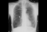

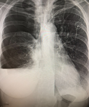
Pneumothorax
Anterior-posterior chest x-ray demonstrating a right pneumothorax
From the collection of Dr Ryland P. Byrd
See this image in context in the following section/s:
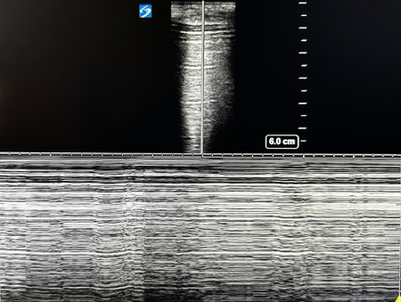
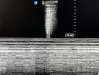
Pneumothorax
POCUS of the lung in a large pneumothorax. Shown is the ultrasound view showing A-lines that are reverberations of the pleural line. There is a lack of B-lines, which is also consistent with a pneumothorax. M-mode is placed on the patient and shows a ‘barcode sign’ confirming presence of air in the pleural space
From the personal collection of Chris Kapp, MD; used with permission
See this image in context in the following section/s:
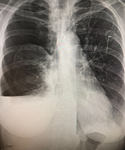
Pneumothorax
Entrapped lung with pneumothorax ex-vacuo. This patient had a malignant pleural effusion that had developed a thick visceral pleural cortical rind preventing re-expansion. Chest tube was for pleural effusion removal, not evacuation of air.
From the personal collection of Chris Kapp, MD; used with permission
See this image in context in the following section/s:
Videos
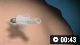 Needle decompression of tension pneumothorax: animated demonstration
How to decompress a tension pneumothorax. Demonstrates insertion of a large-bore intravenous cannula into the fourth intercostal space in an adult.
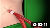 Insertion of intercostal drain, Seldinger technique: animated demonstration
How to insert an intercostal (chest) drain using the Seldinger technique. Video demonstrates: how to identify a safe site for insertion; use of an introducer needle, guidewire, dilators, and intercostal drain; how to confirm drain position; and postprocedure care.
 Insertion of intercostal drain, open technique: animated demonstration
How to insert an intercostal (chest) drain using the open technique. Video demonstrates: tube selection, how to identify the site for drain insertion, how to make the correct incision, how to insert the intercostal drain, how to secure the drain, and postprocedure care.
Use of this content is subject to our disclaimer