History and exam
Key diagnostic factors
common
simultaneous contraction of agonist and antagonist muscles
This is a hallmark of dystonia, but may be better appreciated by electromyography than by clinical examination.
muscle pain
In some cases, pain in the affected muscles may be a prominent feature.
appearance or worsening of dystonia with action
Action dystonia is a near-universal feature, and may be present only with specific tasks involving the dystonic body part or with activation of remote body parts. For example, certain dystonias can be triggered by playing a musical instrument or by writing. When more advanced, dystonia may appear at rest.
blepharospasm
Manifestation of focal dystonia.
cervical torticollis
Manifestation of focal dystonia.
hand spasms
Manifestation of focal dystonia. May be task related.
foot spasms
Manifestation of focal dystonia.
Other diagnostic factors
common
twisting of the affected body part
Twisting of the dystonic body part frequently occurs if the limb, trunk, or neck is involved.
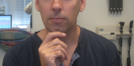
geste antagoniste (sensory trick)
Patients with dystonia are often able to temporarily suppress dystonic posturing or movements by touching the involved region or an adjacent body part. The sensory trick becomes less effective the more severe the dystonia.[Figure caption and citation for the preceding image starts]: Rotational torticollisFrom the personal teaching collections of David K. Simon, MD, Daniel Tarsy, MD, and Ludy C. Shih, MD; used with permissions [Citation ends]. [Figure caption and citation for the preceding image starts]: The torticollis improves with a sensory trick: gently touching his chinFrom the personal teaching collections of David K. Simon, MD, Daniel Tarsy, MD, and Ludy C. Shih, MD; used with permissions [Citation ends].
uncommon
spread to another body part
Focal dystonia may spread to adjacent body parts, becoming segmental or even generalizing over time. This is particularly true with childhood-onset dystonia.
parkinsonism
Accompanying parkinsonism suggests the possibility of Parkinson disease-related dystonia. This usually manifests as foot dystonia, blepharospasm, or cervical dystonia, and rarely as lateral axial dystonia.[36]
myoclonus
Presence of myoclonus as well may indicate a distinct genetic entity called myoclonus-dystonia syndrome.
tremor, weakness, or spasticity
Posttraumatic dystonia is usually accompanied by other neurologic signs including tremor, weakness, and spasticity if head trauma is the cause.
Kayser-Fleischer rings on slit-lamp examination
Presence of Kayser-Fleischer rings suggests Wilson disease.
acute presentation (within 5 days of exposure to antidopaminergic agent)
acute worsening of pre-existing generalized dystonia
Some patients with poorly controlled generalized dystonia may develop acute worsening of their dystonia, which can be severe and life-threatening. Evidence for overlapping neuroleptic malignant syndrome, malignant hyperthermia, serotonin syndrome, or other acute infectious/metabolic/toxic derangement should be considered.[42]
Risk factors
strong
family history of dystonia
A positive family history of dystonia is a significant risk factor, particularly for early-onset cases, and may signify an underlying genetic cause.[13]
A family history is not uniformly present in genetic forms of dystonia due to reduced penetrance.
repetitive activity of affected region
Some forms of focal dystonia are referred to as task-specific dystonia. There is strong anecdotal evidence suggesting that people who frequently use fine motor skills (e.g., musicians), are at increased risk of developing focal dystonia.
birth injury and delayed development in childhood
Documented history of perinatal cerebral injury along with absence of normal early development is more suggestive of an acquired dystonia (cerebral palsy) rather than an idiopathic dystonia.
exposure to antidopaminergic agents
Acute or chronic exposure to both typical and atypical antipsychotic agents and dopamine-receptor blocker antiemetics such as metoclopramide and prochlorperazine are strongly associated with medication-induced dystonias. These can either be acute dystonic reactions or a form of late-onset (tardive) dystonia.
Acute dystonic reactions, most common (but not exclusively) in younger male patients, may include torticollis, tongue or jaw dystonia, oculogyric crisis, or opisthotonus.[30][31][32]
trauma
Posttraumatic dystonia is usually accompanied by other neurologic signs including tremor, weakness, and spasticity if head trauma is the cause. A relatively rapid-onset fixed dystonia accompanied by signs of complex regional pain syndrome is thought to comprise another form of posttraumatic dystonia, usually a focal limb dystonia.[18] This form of dystonia is usually poorly responsive to the treatments used for idiopathic dystonia. There are many reports of patients who have developed what appears to be focal dystonia of a body part that had been traumatically injured days or weeks earlier, although the precise etiologic relationship between trauma and dystonia remains controversial.[19][20][21]
genetic mutation
Several genetic factors have been identified in association with dystonia. These include autosomal-dominant (often with incomplete penetrance), autosomal-recessive, X-linked, and mitochondrial genetic causes.
Mutations in the TOR1A gene (also known as the DYT1 locus) that encodes TorsinA can be found in patients with early-onset dystonia (usually beginning before 26 years of age) that typically begins focally in one limb and subsequently generalizes.[13] Other isolated dystonias can be associated with genetic mutations, including a predominantly neck and upper limb onset dystonia in the second and third decades of life, which is characterized by mutations in the THAP1 gene (also known as the DYT6 locus).[15]
Ashkenazi Jewish ethnicity
The prevalence of dystonia is higher in Ashkenazi Jews, where it is estimated at 1110 per 100,000.[3]
structural lesion of the basal ganglia
parkinsonian syndrome
Some patients with Parkinson disease (PD) or other parkinsonian syndromes can develop dystonia as part of the disease. For example, PD may present with focal limb dystonia, or dystonia may occur in response to levodopa.[3]
Use of this content is subject to our disclaimer