Investigations
1st investigations to order
plain x-rays
Test
Distinguishing acute from chronic fractures on plain radiographs can be difficult. Well-demarcated fracture lines or distinct discontinuity of a thin cortical margin suggest an acute fracture.
An old injury is suggested by sclerosis of the fracture lines, a dense cortical margin, and osteophytes bordering the fracture site.[1][Figure caption and citation for the preceding image starts]: Lateral radiograph showing a T12 compression fracture in osteoporotic bonePersonal collection of Nasir A. Quraishi [Citation ends].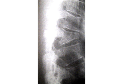
Result
lateral view shows wedging of anterior vertebral body and local kyphosis; anteroposterior view may show interpedicular widening and/or spinous process malalignment suggesting posterior vertebral body involvement
Investigations to consider
CT spine
Test
Useful for assessing bony architecture, but less useful for soft tissues. In patients with new symptomatic vertebral compression fractures identified on radiographs and no known malignancy, CT (or MRI) spine of the area of interest is usually appropriate for pre-procedural planning, if intervention is being considered.[9][50] CT (or MRI) spine of the area of interest is particularly useful for initial imaging if radiographs are negative.[50] CT provides osseous details of axial spine fractures before vertebral augmentation and permits evaluation of vertebral body height, architecture, and integrity of the posterior cortex and pedicles before vertebral augmentation, which is critical in patients with cortical disruption, posterior cortex osseous retropulsion, and spinal canal compression.[9]
Result
shows fracture configuration; useful in excluding unstable crush fractures and pathological fractures
MRI spine
Test
Useful in distinguishing between osteoporotic compression fractures and those caused by underlying tumour or infection.[1][9][50] MRI (or CT) spine of the area of interest is particularly useful for initial imaging if radiographs are negative.[50]
In patients with asymptomatic vertebral compression fractures identified on radiographs and no known malignancy, MRI (or CT) spine of the area of interest is usually appropriate for pre-procedural planning, if intervention is being considered.[9]
Can be helpful for distinguishing healed from recent fractures. This may have predictive value in choosing levels for vertebroplasty.
If there are neurological signs, concerns over fracture stability, or a suspicion that fractures may be pathological, a CT scan and/or an MRI is indicated.[19] MRI is more useful than CT for assessing the integrity of the soft tissue and neural structures, particularly that of the spinal canal.[50] MRI is the only modality for evaluating the internal structure of the spinal cord.[58]
Result
shows fracture with no evidence of soft-tissue mass or signal change extending into the pedicle (which would strongly suggest tumour)
technetium-99m (Tc-99m) whole-body bone scan (bone scintigraphy)
Test
May be helpful to determine the painful vertebrae, particularly the causative level.[9]
Whole-body bone scans may be helpful in the setting of compression fractures to help identify fracture acuity and to appropriately select patients for intervention, particularly if MRI cannot be safely/easily obtained.[50][59]
Result
hot spot at site of fracture
single-photon emission computed tomography (SPECT)/CT
Test
Single-photon emission computed tomography (SPECT) coupled with CT may also be appropriate.[9][50] SPECT imaging provides complementary information because sites of abnormal radio-pharmaceutical uptake on the spine are of interest.[9] SPECT/CT has been shown to localise abnormalities in the vertebra more precisely compared with SPECT imaging alone, particularly in complicated cases, such as multiple collapsed vertebrae of different ages.[9][60] SPECT/CT appears to be comparable to MRI in detecting fractures, particularly in the acute phase, and could be considered if MRI is contraindicated.[61][62]
Result
bone densitometry scan
Test
Confirmatory test for osteoporosis.[11]
Result
shows reduced bone density in osteoporosis
CT myelogram
FBC
Test
A test of exclusion, but may be useful if either malignancy or infection is suspected.
Result
normal
bone profile (including serum calcium and alkaline phosphatase)
Test
A bone profile may be useful to exclude a metabolic cause. This includes serum calcium, albumin, parathyroid hormone, phosphate, alkaline phosphatase, magnesium, creatinine, and serum 25-hydroxyvitamin D (25OHD).
Other tests such as thyroid-stimulating hormone, screening for hypercortisolism, and serum protein electrophoresis may also be considered.
Result
normal
C-reactive protein
Test
A test of exclusion, but may be useful if malignancy or infection is suspected.
Result
normal
blood cultures
Test
A test of exclusion, but may be useful if infection is suspected.
Result
normal
Use of this content is subject to our disclaimer