Aetiology
Vertebral compression fractures in osteoporotic patients are usually due to low-energy trauma in weakened bone. Older adults are more susceptible to spinal injuries related to their increased risk for low velocity falls and the presence of underlying conditions (e.g., osteoporosis and osteopenia, ankylosing spondylitis, and diffuse idiopathic skeletal hyperostosis) which may render the spine highly unstable even after a minor injury.[19] The upper cervical spine is the most common injury location for osteoporotic spinal fractures in older adults.[19][20] These fractures are also most common in patients taking corticosteroid therapy. It is important to rule out a pathological fracture due to tumour infiltration (e.g., multiple myeloma, metastatic disease), metabolic disease (e.g., osteomalacia), or osteomyelitis. See Osteoporosis.
Pathophysiology
Osteoporosis is characterised by decreased bone mass or increased porosity and results in diminished structural support of the bony spinal column.
Type 1 primary osteoporosis: affects postmenopausal women and causes rapid loss of bone after the menopause
Type 2 primary osteoporosis: normally affects patients aged >70 years and causes age-related bone loss.
Secondary osteoporosis is loss of bone due to other diseases or medications (e.g., endocrine disorders, inflammatory changes, or use of corticosteroids) and may occur concurrently with type 1 and type 2 osteoporosis.[21]
The spine is composed mostly of trabecular bone, and osteoporosis is characterised by a decrease in both the size and number of trabeculae. The most frequent site for spinal compression fracture is the thoracolumbar junction, followed by the mid-thoracic spine.
Classification
Denis classification[2]
There is no universally accepted system of classifying osteoporotic spinal compression fractures, but the Denis classification system, based on the 3-column theory of spinal instability, is often used.[3][4]
Anterior column: consists of the anterior two-thirds of the annulus and vertebral body, along with the anterior longitudinal ligament.
Middle column: consists of the posterior one third of the annulus and vertebral body, along with the posterior longitudinal ligament.
Posterior column: consists of all bony and tissue elements posterior to the anterior and middle column.
Radiographic evaluation is used to assess the number of columns involved and likely stability or instability of the fracture.
[Figure caption and citation for the preceding image starts]: Schematic diagram illustrating 3 columns of the Denis classificationCreated by the BMJ Publishing Group [Citation ends].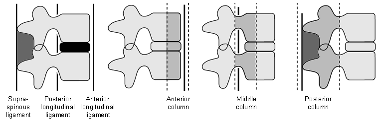
McAfee classification[5]
This classification describes 6 types of spinal fracture based on computed tomography findings and the mode of failure of the middle spinal column.
Wedge compression (isolated anterior column failure)
Stable burst (anterior and middle-column compression failure, posterior column intact)
Unstable burst (compressive failure of anterior and middle columns, disruption of posterior column)
Flexion-distraction (compressive failure of anterior column, tensile failure of posterior column; the centre of rotation is posterior to anterior longitudinal ligament)
Chance fractures (variant of flexion-distraction injury; involves fracture through spinous process, pedicles, and vertebral body)
Translational fractures (disruption of spinal canal alignment in transverse plane, shear mechanism common).
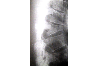
[Figure caption and citation for the preceding image starts]: Lateral radiograph showing a T12 compression fracture in osteoporotic bonePersonal collection of Nasir A. Quraishi [Citation ends].
 [Figure caption and citation for the preceding image starts]: Burst fracture with large retropulsed fragment in spinal canal; the patient underwent posterior decompression and stabilisationPersonal collection of Nasir A. Quraishi [Citation ends].
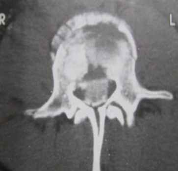 [Figure caption and citation for the preceding image starts]: Vertebral fracture-dislocation; the patient underwent posterior decompression and instrumented stabilisationPersonal collection of Nasir A. Quraishi [Citation ends].
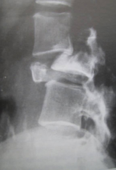
Use of this content is subject to our disclaimer