Images and videos
Images

Uterine fibroids
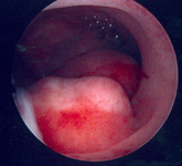
Multiple polyps are identified on hysteroscopic examination of the uterine cavity in this patient with persistent vaginal spotting
From the personal collection of Dr M.F. Mitwally and Dr R.J. Fischer; used with permission
See this image in context in the following section/s:

Uterine fibroids
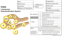
FIGO leiomyoma subclassification system
Munro MG, Critchley HOD, Fraser IS; FIGO Menstrual Disorders Committee. The two FIGO systems for normal and abnormal uterine bleeding symptoms and classification of causes of abnormal uterine bleeding in the reproductive years: 2018 revisions. Int J Gynaecol Obstet. 2018 Dec;143(3):393-408; used with permission.
See this image in context in the following section/s:

Uterine fibroids
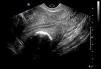
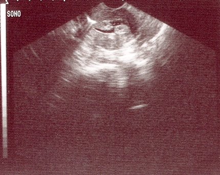
Solitary intramural fibroid as shown by transvaginal pelvic ultrasound (TVUS) demonstrating posterior intramural mass lying between a normal-appearing trilaminar endometrial stripe and posterior uterine serosa
From the personal collection of Dr M.F. Mitwally and Dr R.J. Fischer; used with permission
See this image in context in the following section/s:
Uterine fibroids
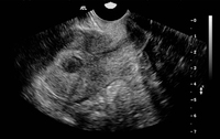
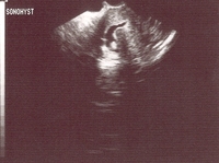
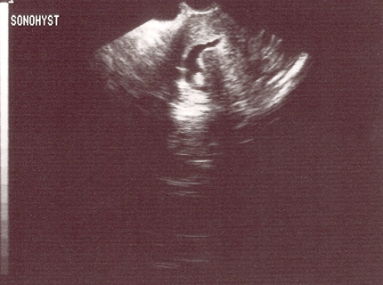
Sonohysterography shows several small intracavitary masses suspicious for polyps
From the personal collection of Dr M.F. Mitwally and Dr R.J. Fischer; used with permission
See this image in context in the following section/s:

Uterine fibroids
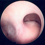
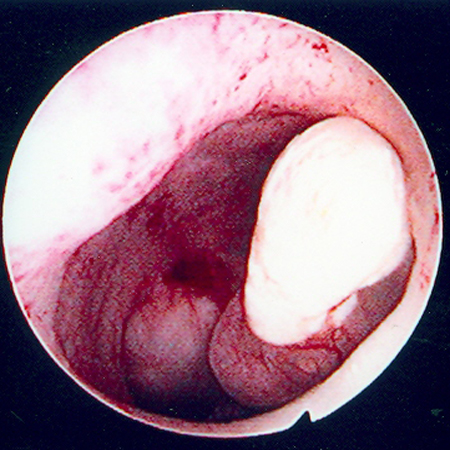
Hysteroscopic image of a large pedunculated submucous uterine fibroid
From the personal collection of Dr M.F. Mitwally and Dr R.J. Fischer; used with permission
See this image in context in the following section/s:
Uterine fibroids
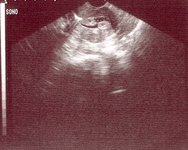
Sonohysterography demonstrates a posterior submucous uterine fibroid deforming the posterior endometrial cavity
From the personal collection of Dr M.F. Mitwally and Dr R.J. Fischer; used with permission
See this image in context in the following section/s:

Uterine fibroids
Hysteroscopic examination of the uterine cavity demonstrates the presence of two contiguous submucous uterine fibroids in this patient with persistent heavy menstrual bleeding
From the personal collection of Dr M.F. Mitwally and Dr R.J. Fischer; used with permission
See this image in context in the following section/s:

Uterine fibroids
Transvaginal pelvic ultrasound (TVUS) shows a midline posterior fundal fibroid greatly distorting the endometrial cavity
From the personal collection of Dr M.F. Mitwally and Dr R.J. Fischer; used with permission
See this image in context in the following section/s:

Uterine fibroids
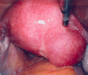
Laparoscopy shows the presence of a large right posterolateral subserosal fibroid; note the close proximity of this uterine fibroid to the right ovary (potential for misdiagnosis as an adnexal mass)
From the personal collection of Dr M.F. Mitwally and Dr R.J. Fischer; used with permission
See this image in context in the following section/s:
Use of this content is subject to our disclaimer