Differentials
Adenomyosis
SIGNS / SYMPTOMS
There are no differentiating signs or symptoms.
INVESTIGATIONS
Distinguished by uterine biopsy and subsequent histopathological examination.
Imaging with pelvic ultrasonography and MRI is also helpful.[79]
Ultrasound sonoelastography is a useful tool to distinguish uterine fibroids and adenomyosis.
Endometrial polyp
SIGNS / SYMPTOMS
There may be no differentiating signs or symptoms. Endometrial polyps do not cause an enlarged uterus.
INVESTIGATIONS
Sonohysterography typically shows a well-circumscribed isoechoic polypoid mass with stalk contained within the endometrial stripe.[46][Figure caption and citation for the preceding image starts]: Multiple polyps are identified on hysteroscopic examination of the uterine cavity in this patient with persistent vaginal spottingFrom the personal collection of Dr M.F. Mitwally and Dr R.J. Fischer; used with permission [Citation ends].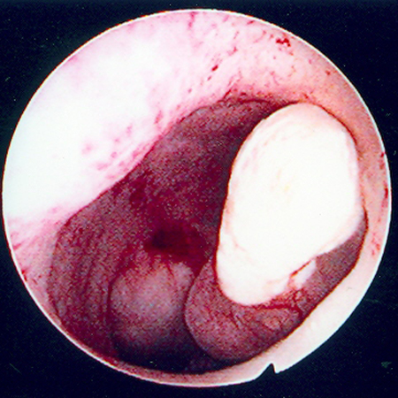
T2-weighted MRI images may show decreased signal intensity compared with endometrium.[65]
Endometrial hyperplasia
SIGNS / SYMPTOMS
There are no differentiating signs or symptoms. Endometrial hyperplasia does not cause uterine enlargement.
INVESTIGATIONS
Endometrial biopsy, and dilatation and curettage, provide differentiation.[80]
Endometrial carcinoma
SIGNS / SYMPTOMS
Because of the high prevalence of uterine fibroids in the general female population, a substantial number of patients with endometrial carcinoma will present with abnormal vaginal bleeding or discharge in association with uterine fibroids.[48]
INVESTIGATIONS
Endometrial sampling: an abnormal endometrial biopsy would show either precursor histology for endometrial carcinoma (simple/complex hyperplasia or simple atypical/complex atypical hyperplasia) or frank endometrial carcinoma.[48]
Dilatation and curettage: persistence of irregular vaginal bleeding despite a negative endometrial biopsy should be pursued by dilatation and curettage.[50]
Uterine sarcoma (leiomyosarcoma, endometrial stromal sarcoma, mixed mesodermal tumour)
SIGNS / SYMPTOMS
Rapid growth of the tumour may be present in uterine sarcomas.
INVESTIGATIONS
No test can reliably diagnose uterine sarcoma.
Serial MRI can identify rapid uterine growth and show characteristics associated with sarcomas such as indistinct borders and invasion into contiguous organs.[65]
Pregnancy
SIGNS / SYMPTOMS
Symptoms of pregnancy (e.g., morning sickness) and missed menstrual period are associated with abdominal expansion over a few weeks.
On examination, uterine enlargement due to pregnancy usually presents as soft, regular, globular pelvic-abdominal mass with its height coinciding with expected gestational age calculated from the last menstrual period.
INVESTIGATIONS
Pelvic ultrasonography visualises the pregnancy sac.[79]
The urine or blood beta-human chorionic gonadotrophin (hCG) pregnancy test is positive. Pregnancy tests usually become positive about 26 to 30 days after the first day of the last menstrual period. Serum beta-hCG test is considered positive if levels are above 5 mIU/mL.
Ovarian cancer
SIGNS / SYMPTOMS
Ovarian cancer is differentiated by rapid tumour growth associated with atypical age for leiomyoma (e.g., post-menopausal women not on hormone replacement therapy), rapid weight loss, or ascites.
INVESTIGATIONS
Pelvic ultrasonography and MRI are useful first-line investigations. MRI may show characteristic low-signal intensity on T2-weighted images seen with uterine fibroids, may show surrounding tissue invasion, and can more exactly define the origin of pelvic masses (uterine versus adnexal).[81]
Surgery and histopathological examination provide differentiation.[79]
Tumours of the gastrointestinal tract and urinary system, lymphomas, and bone tumours
SIGNS / SYMPTOMS
These serious conditions are differentiated by rapid tumour growth associated with atypical age for leiomyoma (e.g., post-menopausal women not on hormone replacement therapy), surrounding tissue invasion, rapid weight loss, or ascites.
INVESTIGATIONS
Pelvic ultrasonography and MRI are useful first-line investigations.
Surgery and histopathological examination provide differentiation.[79]
Use of this content is subject to our disclaimer