Investigations
1st investigations to order
ECG
Test
Perform an ECG as the first line investigation for any patient with suspected Brugada syndrome (BrS).[1][5][7][18][72] Look for type 1, 2, or 3 Brugada patterns, which may indicate BrS.[1]
Type 1 Brugada pattern may be spontaneous or induced (e.g., by factors such as fever or sodium-channel blockers).[1][5][7]
Consider high precordial lead testing if ECG using standard lead placement is not conclusive and you have high clinical suspicion of BrS. High precordial lead testing increases diagnostic yield by accounting for anatomical variations in right ventricular outflow tract anatomy.[1][4][5][65][66][67]
[Figure caption and citation for the preceding image starts]: Electrocardiographic patterns in Brugada syndrome showing type 1 (diagnostic) and types 2 and 3 (non-diagnostic) patterns. Type-1 (diagnostic): coved STT morphology in lead V2 with J-point elevation (dark grey line) of ≥0.2 mV (≥2 mm) and a terminal ST-segment elevation (light grey line, J+60 ms) also ≥0.2 mV (≥2 mm). Note the PR interval and wider QRS complex, wide and deep S in lead I, and fractionation in the right precordial ECG leads. Type-2 (non-diagnostic): saddleback STT morphology in lead V2 with J-point elevation (dark grey line) of ≥0.2 mV (≥2 mm) and a terminal ST-segment elevation (light grey line, J+60 ms) ≥0.1 mV (≥1 mm), followed by a positive T wave. Note the less wide and deep S-wave in lead I, less prominent fractionation. Type-3 (non-diagnostic): saddleback STT morphology in lead V2 with J-point elevation (dark grey line) of ≥0.2 mV (≥2 mm) and a terminal ST-segment elevation (light grey line, J+60 ms) <0.1 mV (<1 mm)Marsman EMJ et al. Heart 2022 May;108(9):668-75; used with permission [Citation ends]. [Figure caption and citation for the preceding image starts]: Standard- and high-lead ECG positions. (Top) Standard-lead ECG positions and corresponding precordial ECG in a patient with Brugada syndrome. (Bottom) High-lead ECG positions and corresponding ECG in the same patient. Note that hV5 and hV6 on the high-lead ECG correspond with V1 and V2 on the standard-lead ECG.Krahn AD, et al. Brugada syndrome. JACC Clin Electrophysiol 2022 Mar;8(3):386-405; used with permission [Citation ends].
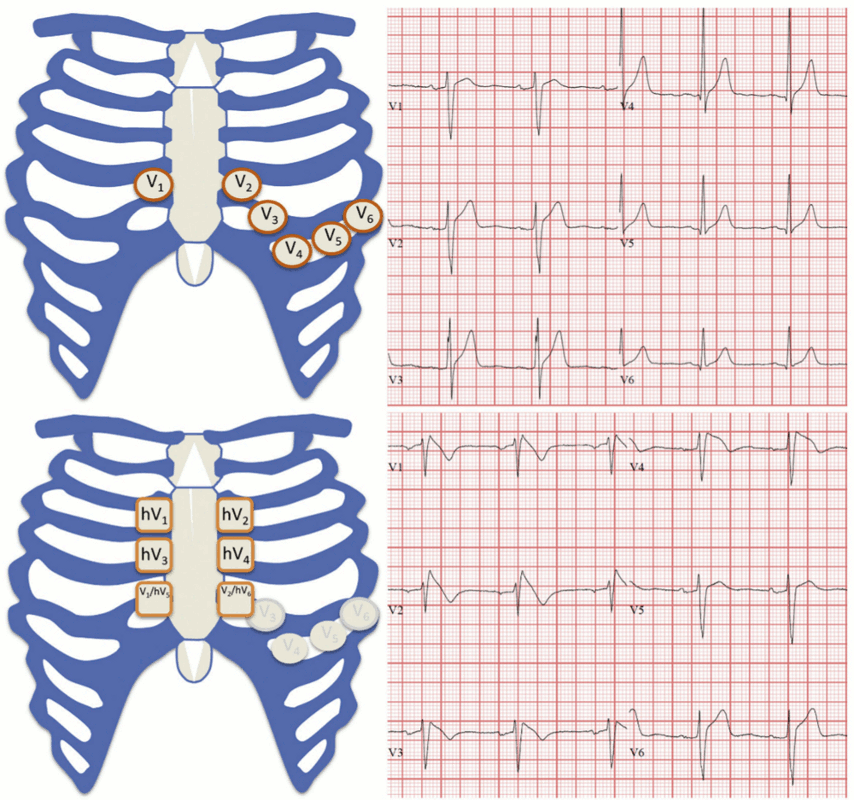
Refer all patients with type 1 Brugada pattern (spontaneous or induced) to a cardiologist or cardiac electrophysiologist.
The diagnosis of BrS is considered probable or definite if spontaneous type 1 Brugada pattern is recorded on ECG from the 2nd to 4th intercostal spaces.[1][4][51][7] However, these patients require diagnostic evaluation by a specialist.
Ensure you establish the patient’s clinical history in all patients with type 1 Brugada pattern.[4] This is essential, because distinguishing type 1 Brugada pattern from other causes of ST elevation using ECG alone can be difficult, even for an experienced cardiologist.[4]
If the patient has induced type 1 Brugada pattern, additional information is key to aid diagnosis of BrS (e.g., relevant clinical history or family history, and/or genetic testing).[1][7]
If the patient has type 2 or 3 Brugada pattern, consider provocative drug testing with sodium channel blockade (see below).[1]
Result
Type 1 Brugada pattern: coved ST-segment elevation (J-point elevation with a gradual down-sloping ST-segment) ≥2 mm with a negative T-wave in the right precordial leads.[1][4][5][7][51] Type 2 or 3 Brugada pattern: saddleback ST-segment configuration with variable levels of ST-segment elevation.[1][4][51]
echocardiogram
Test
Organise an echocardiogram for any patient being evaluated for Brugada syndrome (BrS) to assess for underlying structural heart disease and to rule out other causes of their presentation.[1][49]
Result
normal; mild structural abnormalities in the right ventricle or right ventricular outflow tract
Investigations to consider
provocative drug testing with sodium channel blockade
Test
Consider provocative drug testing with sodium-channel blockers (e.g., procainamide, flecainide) for any patient with both:[1][7][13][49]
Type 2 or 3 Brugada pattern (Brugada pattern) on
AND
Suspected BrS due to relevant clinical signs, symptoms, or family history.
The diagnosis of BrS is considered probable if type 1 Brugada pattern is provoked on ECG during drug testing with sodium-channel blockers; this scores 2 points on the Shanghai score (see Criteria).[1] Refer these patients to a cardiologist or cardiac electrophysiologist.
Due to an associated risk of inducing life-threatening ventricular arrhythmias, provocative drug testing should only be performed by experts under optimal circumstances.[68]
Sodium channel blocker testing is not recommended in patients with a prior type I Brugada pattern.[7]
genetic testing for BrS
Test
Arrange genetic testing in patients with type 1 Brugada ECG pattern (spontaneous or induced); this helps facilitate family screening of first degree relatives.[1][18][69] However, the presence of a susceptible gene mutation is not diagnostic of BrS. This is in part due to only 50% penetrance of genetic variants. Clinical presentation remains central to diagnosing Brugada syndrome.[1] Probable pathogenic mutation in a BrS susceptibility gene also scores 0.5 points on the Shanghai score (see Criteria).[1]
Mutations of many genes have been implicated in BrS, but only SCN5A gene variants are considered definitely disease-causing.[1][7] However, identifiable SCN5A variants are only found in approximately 20% to 30% of patients with BrS.[1][7][16][17][18][72] Mutations of other genes only account for around 2% to 5% of cases.[14][16][17][19]
Result
positive for known pathogenic mutation associated with Brugada syndrome (e.g., SCN5A)
advanced cardiac imaging (MRI or CT)
Test
Consider other advanced imaging modalities, such as cardiac MRI or CT if the diagnosis of Brugada syndrome (BrS) is uncertain to help differentiate BrS from other differentials, such as arrhythmogenic cardiomyopathy.[36][70] See Differentials.
Result
may demonstrate cardiac structural changes, particularly in the right ventricular outflow tract
invasive electrophysiological (EP) study with inducibility testing for ventricular arrhythmias
Test
Invasive EP studies (including measurement of baseline intervals, programmed electrical stimulation [PES], and electroanatomical mapping) with inducibility testing may be considered following consultation with an electrophysiologist or cardiologist with expertise in managing BrS if the patient has asymptomatic Brugada syndrome (BrS) and is deemed intermediate risk on risk stratification - see Management approach for more information about risk stratification.[7][72] Although there is debate regarding the prognostic value of PES in primary electrical diseases, there is some evidence to consider its use in BrS.[72][71]
Invasive EP assessments have demonstrated voltage abnormalities recorded from the right ventricular epicardium of patients with BrS in the absence of cardiac MR or CT structural abnormalities.[38] This highlights the importance of expert consultation if you suspect BrS, but the diagnosis is unclear.
Result
inducible ventricular tachycardia or fibrillation in patients with asymptomatic BrS
Use of this content is subject to our disclaimer