Images and videos
Images


Pericarditis
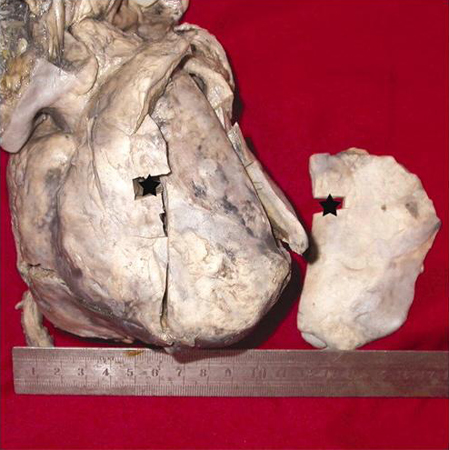
Constrictive pericarditis identified at autopsy; the right section is scar tissue cut from the very front of the heart
Xu JD, Cao XX, Liu XP, et al. BMJ Case Reports 2009; doi:10.1136/bcr.03.2009.1688
See this image in context in the following section/s:

Pericarditis
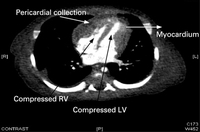
Chest CT in a baby with purulent pericarditis, showing a pericardial collection with compression of the left (LV) and right (RV) ventricles
Karuppaswamy V, Shauq A, Alphonso N. BMJ Case Reports 2009; doi:10.1136/bcr.2007.136564
See this image in context in the following section/s:
Pericarditis
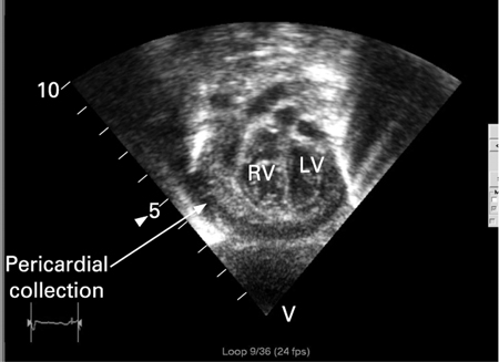
Echocardiogram in a baby with purulent pericarditis, showing a pericardial collection. LV = left ventricle, RV = right ventricle
Karuppaswamy V, Shauq A, Alphonso N. BMJ Case Reports 2009; doi:10.1136/bcr.2007.136564
See this image in context in the following section/s:
Pericarditis
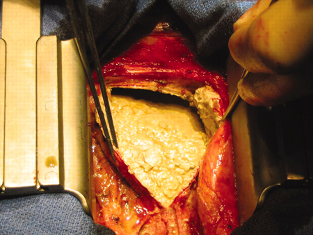
Pericardiectomy in a 56-year-old male patient with idiopathic calcific constrictive pericarditis. The pericardium is thickened and calcified
Patanwala I, Crilley J, Trewby PN. BMJ Case Reports 2009; doi:10.1136/bcr.06.2008.0015
See this image in context in the following section/s:
Pericarditis
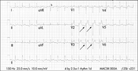
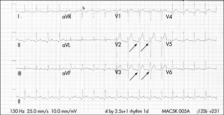
ECG in a patient with acute pericarditis, showing diffuse ST-segment elevation in the precordial leads. There is also PR-segment depression in leads V2-V6 (arrows)
Rathore S, Dodds PA. BMJ Case Reports 2009; doi:10.1136/bcr.2006.097071
See this image in context in the following section/s:

Pericarditis
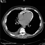
Chest CT showing a double layer of pericardial calcification in a 56-year-old male patient with idiopathic calcific constrictive pericarditis
Patanwala I, Crilley J, Trewby PN. BMJ Case Reports 2009; doi:10.1136/bcr.06.2008.0015
See this image in context in the following section/s:
Use of this content is subject to our disclaimer