Images and videos
Images

Established atrial fibrillation
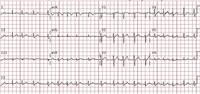
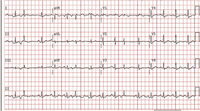
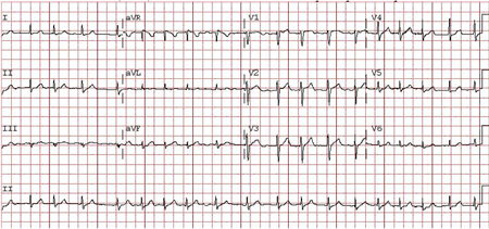
Atrial tachycardia: bursts of atrial tachycardia (10 beats in the middle section of the rhythm strip II at bottom) follows sinus complexes
From the collection of Dr Arti N. Shah
See this image in context in the following section/s:
Established atrial fibrillation
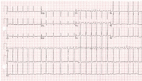
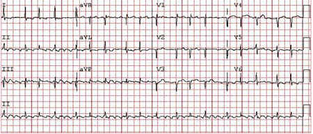
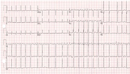
Atrial flutter: typical saw-tooth appearance of the flutter waves in the inferior leads (leads II, III, and aVF) indicates typical counterclockwise atrial flutter; the ventricular (QRS complexes) rate is variable
From the collection of Dr Arti N. Shah
See this image in context in the following section/s:
Established atrial fibrillation
Atrial fibrillation: P waves are not discernible; the ventricular (QRS complexes) rate is irregularly irregular
From the collection of Dr Arti N. Shah
See this image in context in the following section/s:

Established atrial fibrillation
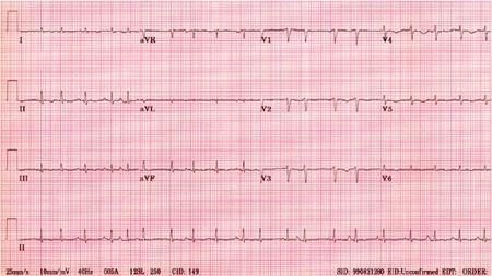
Sinus rhythm with premature atrial complexes
From the collection of Dr Arti N. Shah
See this image in context in the following section/s:
Established atrial fibrillation
Sinus rhythm with premature ventricular complexes
From the collection of Dr Arti N. Shah
See this image in context in the following section/s:
Established atrial fibrillation
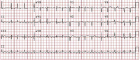
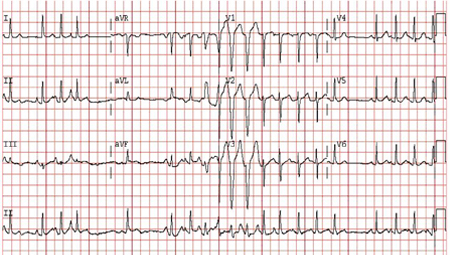
Atrial fibrillation: P waves are not discernible; 'fibrillatory or f waves' of varying amplitudes and very rapid and irregular rates are particularly seen in lead V1; the ventricular (QRS complexes) rate is irregularly irregular
From the collection of Dr Bharat K. Kantharia
See this image in context in the following section/s:
Established atrial fibrillation
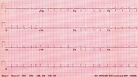
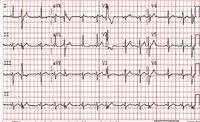
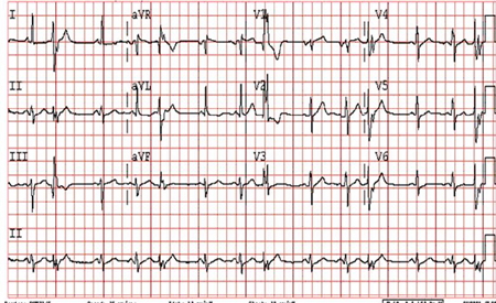
Multifocal atrial tachycardia: there are P waves of multiple (at least 3) different morphologies
From the collection of Dr Arti N. Shah
See this image in context in the following section/s:
Videos
 How to perform an ECG animated demonstration
How to record an ECG. Demonstrates placement of chest and limb electrodes.
Use of this content is subject to our disclaimer