This page compiles our content related to skin cancer. For further information on diagnosis and treatment, follow the links below to our full BMJ Best Practice topics on the relevant conditions and symptoms.
Introduction
Relevant conditions
Merkel cell carcinoma (MCC)Signs & symptomsInvestigationsDifferentialsTreatment algorithm | go to our full topic on Merkel cell carcinoma (MCC) MCC is a rare, aggressive cutaneous neuroendocrine neoplasm that primarily affects white adults. Incidence increases with age and is highest among those aged >85 years.[5] Two distinct aetiologies are recognised: exposure to ultraviolet radiation and oncogenic transformation by Merkel cell polyomavirus. Diagnosis is confirmed with skin biopsy and histopathological evaluation including immunohistochemistry. Occult metastasis is common, and sentinel lymph node biopsy and imaging are important tools for staging the disease. |
|---|---|
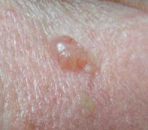
Basal cell carcinomaSigns & symptomsInvestigationsDifferentialsTreatment algorithm | go to our full topic on Basal cell carcinoma The most common malignancy of the skin in fair-skinned adults in the US, Australia, and Europe.[6]
CRUK Cancer incidence statistics
Opens in new window It typically presents as pearly papules and/or plaques; non-healing scabs; small crusts and non-healing wounds; plaques, nodules, and tumours with rolled borders; or papules with associated telangiectasias.[7][8][Figure caption and citation for the preceding image starts]: Nodular basal cell carcinoma on the cheek, on background of diffuse solar damage with marked solar elastosisFrom the collection of Dr Robert A. Schwartz [Citation ends]. |
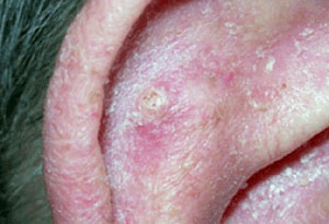
Squamous cell carcinomaSigns & symptomsInvestigationsDifferentialsTreatment algorithm | go to our full topic on Squamous cell carcinoma Ranges from in situ tumours (Bowen's disease) to invasive tumours and metastatic disease. Patients commonly present with a new or enlarging lesion that they are concerned about, which may be tender or itchy, or a non-healing wound originally caused by some trauma. In situ tumours are typically thin, flesh-coloured to erythematous, scaly plaques, while invasive squamous cell carcinoma (SCC) may present as an exophytic tumour or ulcer.[Figure caption and citation for the preceding image starts]: Squamous cell carcinoma on the ear with surrounding actinic damageFrom the collection of Dr Jessica M. Sheehan and Dr Keyoumars Soltani [Citation ends]. |
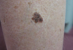
MelanomaSigns & symptomsInvestigationsDifferentialsTreatment algorithm | go to our full topic on Melanoma A malignant tumour arising from pigment-producing melanocytes found in the skin, eye, and central nervous system. Several variants exist. Typically presents as a deeply pigmented skin lesion that is new or changing in size, shape, or colour.[10][11][Figure caption and citation for the preceding image starts]: Superficial spreading malignant melanomaFrom the collection of Dr Hobart W. Walling [Citation ends]. |
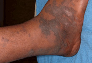
Kaposi's sarcomaSigns & symptomsInvestigationsDifferentialsTreatment algorithm | go to our full topic on Kaposi's sarcoma A low-grade vasoformative neoplasm associated with human herpesvirus-8 (HHV-8, also knows as Kaposi's sarcoma-associated herpesvirus [KSHV]) infection.[16] Lesions frequently involve mucocutaneous sites, but may become more extensive to involve the lymph nodes and visceral organs. Skin lesions evolve from an early patch, to a plaque, and later to ulcerating tumour nodules.[Figure caption and citation for the preceding image starts]: Kaposi sarcoma cutaneous purple-brown plaque on the footFrom the collection of Dr Bruce J. Dezube [Citation ends]. |
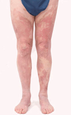
Cutaneous T-cell lymphomaSigns & symptomsInvestigationsDifferentialsTreatment algorithm | go to our full topic on Cutaneous T-cell lymphoma Heterogeneous group of uncommon disorders characterised by clonal accumulation of T lymphocytes primarily or exclusively in the skin. Mycosis fungoides and its leukaemic variant, Sézary syndrome, are the most common subtypes.[18] Establishing a diagnosis is often difficult, as the disease can manifest in a number of different ways, including flat patches, raised plaques, large tumours, and/or marked erythroderma (intense and widespread reddening of the skin).[Figure caption and citation for the preceding image starts]: Cutaneous T-cell lymphoma: extensive patch diseaseFrom the collection of the Christie NHS Foundation Trust, Manchester, UK; used with permission [Citation ends]. |
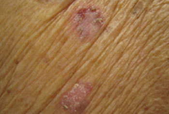
Actinic keratosisSigns & symptomsInvestigationsDifferentialsTreatment algorithm | go to our full topic on Actinic keratosis Lesions are skin-coloured, yellowish, or erythematous, ill-defined, irregularly shaped, small, scaly macules or plaques localised in sun-exposed areas of the body (e.g., forehead, lower lip, dorsum of the hands, forearms, bald areas of the scalp, and ears).[Figure caption and citation for the preceding image starts]: Regular actinic keratosisFrom the collection of the Department of Dermatology and Cutaneous Surgery, University of Miami Miller School of Medicine [Citation ends]. |
SunburnSigns & symptomsInvestigationsDifferentialsTreatment algorithm | go to our full topic on Sunburn Sunburn is an acute inflammatory reaction of the skin induced by overexposure to UV radiation. Skin findings include erythema and oedema, with or without vesiculation, followed by desquamation. Symptoms include pain and/or pruritus. Acute sunburn is a self-limiting condition and typically requires only supportive care. Primary prevention via sun avoidance, physical protection, and the appropriate use of sunscreen is key to managing the condition, as cellular damage caused by UV radiation is irreversible and may with time increase the risk of skin cancer. |
Contributors
Authors
Editorial Team
BMJ Publishing Group
Disclosures
This overview has been compiled using the information in existing sub-topics.
Use of this content is subject to our disclaimer