History and exam
Key diagnostic factors
common
presence of risk factors
firm, non-tender, red or pink-to-violaceous or skin-coloured papule or subcutaneous nodule
The primary cutaneous MCC tumour usually presents as a solitary, firm, erythematous or pink-to-violaceous or skin-coloured papule or subcutaneous nodule, typically on sun-exposed skin.[7][19]
MCC lesions most frequently develop on the sun-exposed areas of head and neck (29% to 44%) and the extremities (37% to 45%). Less than 10% of MCCs occur on partially sun-protected areas (trunk, thighs, and hair-bearing scalp) or highly sun-protected areas (buttocks). Extra-cutaneous sites (e.g., vulva, vagina, oral mucosa, parotid gland, nasal cavity) are very rarely involved.
MCC lesions are typically asymptomatic and not tender to the touch.[7] One study of 195 patients with pathologically confirmed MCC found that 88% of tumours were asymptomatic.[2]
[Figure caption and citation for the preceding image starts]: A primary MCC tumour in an 87-year-old man who presented with a large, fast-growing, red, eroded nodule on the right templeFrom the collection of Dr Kelly Harms and Dr Alison Lee, University of Michigan; used with patient consent [Citation ends].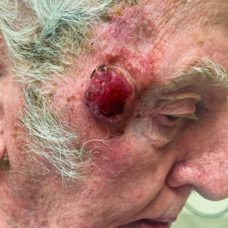 [Figure caption and citation for the preceding image starts]: MCC on the left lower lip of a 68-year-old man. The asymptomatic tumour is an ill-defined reddish-pink, scaly plaque blurring the lower vermillion of the lipFrom the collection of Dr Kelly Harms and Dr Alison Lee, University of Michigan; used with patient consent [Citation ends].
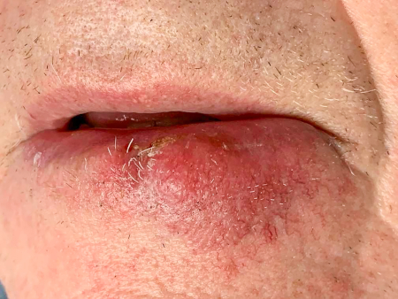 [Figure caption and citation for the preceding image starts]: A 2 cm red, nodular MCC with varied vessel patterns on the right upper arm of an 80-year-old manFrom the collection of Dr Kelly Harms and Dr Alison Lee, University of Michigan; used with patient consent [Citation ends].
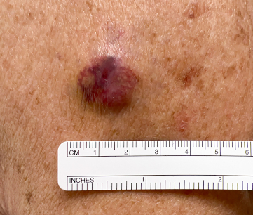
For further clinical images of MCC, see Diagnosis approach.
rapidly growing lesion
The primary MCC tumour typically grows rapidly over the first 3 months.[2]
Other diagnostic factors
common
uncommon
ulceration or bleeding cutaneous lesion
small papules or nodules surrounding a primary lesion
Cutaneous satellite or in-transit metastases present as smaller papules or nodules surrounding the primary tumour.
Risk factors
strong
cumulative ultraviolet (UV) exposure
UV-mediated DNA damage is a significant risk factor for MCC.
MCC is strongly associated with lower latitudes and high UV radiation indexes.[13] The primary tumour typically occurs on sun-exposed skin.[2][7]
MCC tumours that do not harbour integration of Merkel cell polyomavirus (MCPyV) demonstrate a high burden of UV-signature mutations, and UV-mediated DNA damage is considered a pathogenic mechanism of MCC tumorigenesis.[22][23]
immunosuppression
MCC is strongly associated with immunosuppression, with an estimated 6% to 12% of all patients with MCC being immunosuppressed.[7][19]
There is a greater than 34-fold increased risk of MCC in patients with chronic lymphocytic leukaemia (CLL).[15][17] Patients with solid organ transplantation have a 23% increased risk of MCC.[16][27] Other immunosuppression mechanisms, such as human immunodeficiency virus (HIV) infection and use of immunosuppressive medications, have also been associated with a higher risk of developing MCC.[7][28]
The European Society for Medical Oncology (ESMO) guideline states that 10% of patients with MCC are organ transplant recipients, or have either a haematological malignancy or HIV infection.[19]
advancing age
male sex
white skin
Merkel cell polyomavirus (MCPyV) infection with oncogenic transformation
Oncogenic transformation by MCPyV is a key risk factor for MCC.[29] MCPyV is a ubiquitous virus, with seroprevalence in the adult population of 60% to 80%.[20] Oncogenic transformation by MCPyV is a rare event and requires viral integration into the host genome, resulting in inactivation of a viral replication protein. MCPyV is associated with up to 70% to 80% of MCC tumours in North America and Europe, with lower associations in Australia, where a higher proportion of cases are associated with UV exposure.[7][12][14]
Use of this content is subject to our disclaimer