Investigations
1st investigations to order
biopsy
Test
SCC in situ (Bowen's disease) displays full-thickness atypia that is confined to the epidermis with an intact basement membrane.[Figure caption and citation for the preceding image starts]: Biopsy showing histology of an in-situ SCC with full-thickness keratinocyte atypiaFrom the private collection of Dr Nwaneshiudu and Dr Soltani [Citation ends].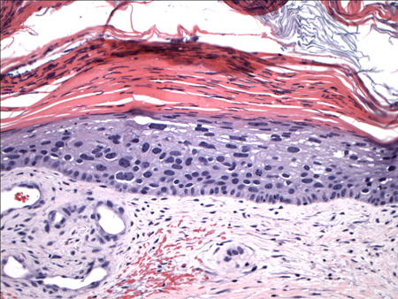
Invasive tumours extend beyond the basement membrane, penetrate into the dermis, and may invade deeper structures.[Figure caption and citation for the preceding image starts]: Biopsy histology showing invasive SCC with keratinocyte atypia involving all layers of the epidermis and islands budding into the dermisFrom the private collection of Dr Nwaneshiudu and Dr Soltani [Citation ends].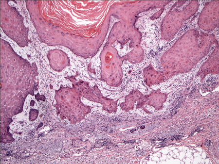
Keratoacanthomas appear as endophytic nodules with a central invagination, replete with keratin. Atypia is minimal and mitoses are rare. Lymphocytic infiltrate is present at the margins of the lesion.
Marjolin ulcers appear as SCC.
Verrucous carcinoma appears as a well-differentiated neoplasm with warty features such as acanthosis, hyperkeratosis, and hypergranulosis.
Actinic keratoses show histological changes of intraepidermal keratinocytic dysplasia, especially in the basal layer.
Result
full thickness keratinocyte atypia
Investigations to consider
CT scan
Test
For high-risk squamous cell carcinoma (i.e., larger and more invasive lesions), perineural invasion, or lymphadenopathy, a CT scan of the area of interest may be ordered to evaluate the extent of the large tumours and/or to rule out metastatic disease.[75] Some evidence suggests that CT is the more accurate modality to detect bony invasion and nodal metastasis.[77]
Result
lymphadenopathy and/or visceral nodules suggestive of metastases
MRI scan
Test
For high-risk squamous cell carcinoma (i.e., larger and more invasive lesions), perineural invasion, or lymphadenopathy, an MRI scan of the area of interest may be ordered to evaluate the extent of the large tumours and/or to rule out metastatic disease.[75] Some evidence suggests that MRI is more accurate than CT for the detection of perineural spread.[77]
Result
lymphadenopathy and/or visceral nodules suggestive of metastases
PET scan
Test
For high-risk squamous cell carcinoma (i.e., larger and more invasive lesions), perineural invasion, or lymphadenopathy, a PET scan of the area of interest may be ordered to evaluate the extent of the large tumours and/or to rule out metastatic disease.[75]
Result
lymphadenopathy and/or visceral nodules suggestive of metastases
FBC with differential
Test
For high-risk squamous cell carcinoma (i.e., larger and more invasive lesions), to facilitate early detection of metastases.
Secondary to bone marrow involvement.
Result
normal, except if bone marrow metastases present
LFTs
Test
For high-risk squamous cell carcinoma (i.e., larger and more invasive lesions), to facilitate early detection of metastases.
Result
normal, except if liver metastases present
Use of this content is subject to our disclaimer