Summary
Definition
History and exam
Key diagnostic factors
- presence of risk factors
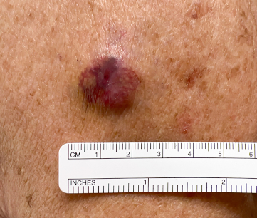
- firm, non-tender, red or pink-to-violaceous or skin-coloured papule or subcutaneous nodule
- rapidly growing lesion
Other diagnostic factors
- enlarged lymph nodes
- ulceration or bleeding cutaneous lesion
- small papules or nodules surrounding a primary lesion
Risk factors
- cumulative ultraviolet (UV) exposure
- immunosuppression
- advancing age
- male sex
- white skin
- Merkel cell polyomavirus (MCPyV) infection with oncogenic transformation
Diagnostic investigations
Investigations to consider
- lymph node ultrasound
- whole-body PET scan
- CT scan with contrast chest/abdomen/pelvis (± neck)
- brain MRI
- fine needle aspiration or core biopsy
- sentinel lymph node biopsy (SLNB)
Treatment algorithm
Contributors
Authors
Kelly Harms, MD, PhD
Associate Professor of Dermatology
Michigan Medicine
University of Michigan
Ann Arbor
MI
Disclosures
KH is a panel member for NCCN guidelines on Merkel cell carcinoma. She serves as Chair of Professionalism and Ethics for the Michigan Dermatological Society Board of Directors. KH is married to Paul Harms, a co-author of research on Merkel cell carcinoma.
Alison Lee, MD, MHS
Clinical Fellow
Department of Dermatology
Michigan Medicine
University of Michigan
Ann Arbor
MI
Disclosures
AL declares that she has no competing interests.
Peer reviewers
Isaac Brownell, MD, PhD
Senior Investigator
Dermatology Branch
National Institute of Arthritis and Musculoskeletal and Skin Diseases (NIAMS)
National Institutes of Health (NIH)
Bethesda
MD
Disclosures
IB declares that he has no competing interests.
Zoe Venables, MbChB, MMedSci, MRCP
Clinical Associate Professor and Consultant Dermatologist
Norfolk and Norwich University Hospitals NHS Foundation Trust
Norwich
UK
Disclosures
ZV declares that she has no competing interests.
Use of this content is subject to our disclaimer