Images and videos
Images


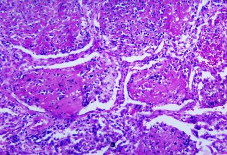
Histoplasmosis
Histopathologic changes associated with histoplasmosis of the lung
Dr Martin Hicklin, Public Health Image Library, US Centers for Disease Control and Prevention
See this image in context in the following section/s:
Histoplasmosis
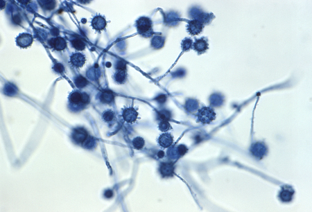
Histoplasma capsulatum in a soil sample
Dr Libero Ajello, Public Health Image Library, US Centers for Disease Control and Prevention
See this image in context in the following section/s:

Histoplasmosis
Gross pathology specimen of lung showing cut surface of fibrocaseous nodule due to Histoplasma capsulatum
Public Health Image Library, US Centers for Disease Control and Prevention and ASCP Atlas of Clinical Mycology II
See this image in context in the following section/s:

Histoplasmosis
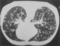
Lung CT scan showing classic "snowstorm" appearance of acute histoplasmosis
Public Health Image Library, US Centers for Disease Control and Prevention
See this image in context in the following section/s:

Histoplasmosis
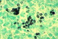
Methenamine silver stain revealing Histoplasma capsulatum fungi in lung tissue
Dr Edwin P. Ewing, Jr., Public Health Image Library, US Centers for Disease Control and Prevention
See this image in context in the following section/s:

Histoplasmosis
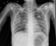
Chest x-ray of a patient with histoplasmosis, demonstrating bilateral diffuse reticulonodular infiltrates
From the personal collection of Dr David L. Goldman
See this image in context in the following section/s:

Histoplasmosis
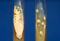
Slant cultures growing Histoplasma capsulatum colonies on 2 different kinds of agar
Dr Lenore Haley, Public Health Image Library, US Centers for Disease Control and Prevention
See this image in context in the following section/s:
Use of this content is subject to our disclaimer