History and exam
Key diagnostic factors
common
bilateral cleft lip ± palate
Most commonly presents with a complete bilateral cleft palate. However, incomplete bilateral and even unilateral cleft palate is also seen. [Figure caption and citation for the preceding image starts]: Bilateral cleft lip and palate preoperativelyFrom the collection of Travis T. Tollefson, MD, FACS [Citation ends].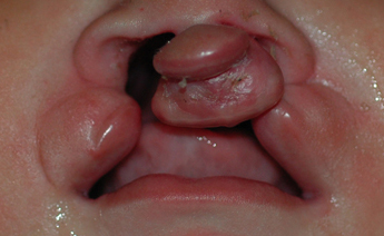
The configuration of the lateral segments of a bilateral cleft lip with or without cleft palate is similar to that of the lateral segment of the unilateral deformity.
The central prolabium and premaxilla in bilateral cleft lip and palate are markedly different from those of the unilateral deformity, as there is no muscle in the prolabial skin due to its embryologic origin.
Varying severity of clefting of the lip, alveolus, and palate are observed. In complete bilateral cleft lip with or without cleft palate, the premaxilla protrudes anteriorly and is totally detached from each maxilla. In incomplete bilateral cleft lip, there is usually some skeletal continuity and very little protrusion of the premaxilla and prolabium. If the lateral maxillary alveolar segments are constricted together, a "locked-out" premaxilla is noted. The complete bilateral cleft lip nasal columella is short, contributing to bilateral hooding of the ala and a wide, bulbous deprojected nasal tip.
unilateral cleft lip ± palate
There is extensive variation in severity from complete to microform. [Figure caption and citation for the preceding image starts]: Unilateral cleft lip and palate preoperativelyFrom the collection of Travis T. Tollefson, MD, FACS [Citation ends].
In severely wide (>1 cm) complete unilateral cleft lip and palate, there is complete separation of the lip musculature, alveolus, and palate.
A microform cleft is the most diminutive form of lip cleft.
An incomplete cleft lip with or without cleft palate extends to more than a quarter of the labial height, and is measured from the normal peak of the upper lip junction between the white and red lip (Cupid's bow) to the bottom of the nostril (nasal sill).
In an isolated unilateral cleft lip, the base of the nose splays laterally when the infant smiles. The nose and lip on the noncleft side is characterized by a short vertical white lip height, deficient medial mucosa, deficient columellar skin, caudal septal deflection to the noncleft side, and a cleft lip nasal deformity with an asymmetric nasal tip (secondary to lower lateral cartilage dysmorphology).
isolated cleft palate
Degree of clefting can range from a complete isolated cleft palate to a bifid uvula.
An example of a deformational cleft palate is seen in Pierre Robin sequence, which is characterized by the classic triad of micrognathia, glossoptosis, and an isolated cleft palate. [Figure caption and citation for the preceding image starts]: Lateral view of infant with Pierre Robin sequenceFrom the collection of Travis T. Tollefson, MD, FACS [Citation ends].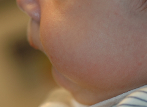
microform cleft lip
Characterized by dysmorphic features including a minor nasal deformity, philtral groove, indented free mucosal margin, and notched vermilion-cutaneous junction with disruption extending to no more than a quarter of the labial height, measured from the normal peak of the upper lip junction between the white and red lip (Cupid's bow) to the bottom of the nostril (nasal sill).[39][Figure caption and citation for the preceding image starts]: Microform cleft lipFrom the collection of Travis T. Tollefson, MD, FACS [Citation ends].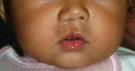
isolated submucous cleft palate
Less overt palatal clefting may be missed on examination and may instead present with symptoms of feeding difficulties, airway obstruction, and/or poor weight gain.
Identified by a diastasis of the midline palatal musculature (zona pellucida), a notched hard palate on palpation, and a bifid uvula. [Figure caption and citation for the preceding image starts]: Submucous cleft palateFrom the collection of Travis T. Tollefson, MD, FACS [Citation ends].
May also present with hypernasal speech or nasal regurgitation.
positive prenatal ultrasound
Routine scanning at approximately 18 weeks' gestation can identify the possible presence of a cleft lip.
Other diagnostic factors
common
difficulty feeding
Less overt palatal clefting may be missed on examination and may instead present with symptoms of feeding difficulties. Breastfeeding is possible with an isolated cleft lip. However, neonates with a cleft palate often cannot produce the negative pressures necessary for suction.
poor weight gain
Less overt palatal clefting may be missed on examination and may instead present with symptoms of poor weight gain secondary to feeding difficulties.
uncommon
airway obstruction
A small proportion of infants with any type of cleft palate, including Pierre Robin sequence (triad of cleft palate, microgenia, and glossoptosis), will present with symptoms requiring immediate airway management.
Less overt palatal clefting may be missed on examination and may instead present with symptoms of airway obstruction.
Risk factors
strong
genetic predisposition
Includes major chromosomal abnormalities associated with many genetic syndromes and inherited, nonsyndromic clefts in families with affected first-degree relatives.
Studies show that monozygotic twins have 43% paired concordance, compared to 5% in dizygotic twins.[13] Parents without clefts have a 4% recurrence rate in future pregnancies following the birth of 1 affected child.[14] However, the risk for a third child after having 2 affected children rises from 4.4% to 9%.[15] A parent with cleft lip and/or palate has a 4% risk of having an affected child. If further first-degree relatives are affected by a cleft palate, the risk to the child rises to between 10% and 20%.[14]
Van der Woude syndrome is the most common syndromic cause of orofacial clefting, accounting for 2% of cases.[7] It is caused by a mutation in the interferon regulatory factor 6 (IRF6) gene and is characterized by cleft lip +/- cleft palate, lip pits, skin folds, syndactyly, and oral adhesions.[11][Figure caption and citation for the preceding image starts]: Repaired bilateral cleft lip showing the lip pits of van der Woude syndromeFrom the collection of Travis T. Tollefson, MD, FACS [Citation ends].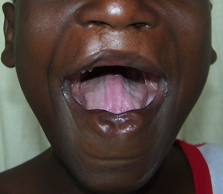
anticonvulsant drugs
Phenytoin and other anticonvulsant medications taken during pregnancy have been linked to a number of congenital deformities, including cleft lip and palate.[20]
maternal tobacco use
Although the pathophysiology behind the association is not yet understood, the risk of orofacial clefting is increased in mothers who use tobacco during pregnancy or periconceptionally (1 month before and 3 months after conception). There are a number of meta-analyses that show a statistically significant, dose-dependent link between maternal smoking and cleft lip with or without cleft palate and isolated cleft palate.[26][27][28][29][30]
weak
maternal alcohol consumption
Higher quantities of maternal alcohol consumption (>5 units per drinking session) are associated with an increased incidence of orofacial clefts in women with particular alcohol dehydrogenase gene variants.[31]
folic acid deficiency
Conflicting reports have been published regarding the efficacy of prenatal folic acid supplementation to reduce the risk of orofacial clefting. Some studies show a decreased risk of clefting, while others show no reduction in the incidence of orofacial clefts.[35][22] A Cochrane review of randomized controlled trials showed that prenatal folic acid had an overall protective effect against neural tube defects. However, no relation to decreased cleft lip and palate was found.[21][23] Folic acid, alone or in combination with vitamins and minerals, prevents neural tube defects but does not have a clear effect on other birth defects.
Maternal perinatal multivitamin use has been shown to be more protective against cleft lip and palate in infants with certain transforming growth factor-alpha (TGF-A) genotypes.[24] It has been shown that recurrence rates for clefting (i.e., the incidence of clefting if one sibling has already been born with an orofacial cleft) is decreased significantly with the use of folic acid, although no dose-dependent relationship has been established.[25]
Use of this content is subject to our disclaimer