Emerging treatments
Recombinant bone morphogenetic protein
The alveolar cleft is traditionally treated, after orthodontic preparation between the ages of 8 and 11 years, with an alveolar bone graft of cancellous iliac crest bone, but use of a tissue-engineered bone growth factor such as recombinant bone morphogenetic protein-2 (BMP-2) is a promising alternative.[66] Long-term outcomes in alveolar cleft grafting are not yet available.
Gingivoperiosteoplasty (GPP)
There are opposing opinions regarding the use of GPP that is performed after the alveolar segments have been approximated with presurgical nasal alveolar molding (PNAM). Although GPP is thought to inhibit maxillary bone growth and lead to inadequate alveolar bone growth for orthodontia or dental implantation, it prevents the need for an alveolar bone graft in 60% of patients (thus reducing health care costs).[51][67][68] Maxillary bone growth disturbance may be reduced with a limited mucoperiosteal dissection.[55] There are currently no studies reporting long-term maxillary growth outcomes with GPP and limited mucoperiosteal dissection.
Primary rhinoplasty at the time of lip repair
Although cleft lip outcomes have improved, the stigma of cleft lip nasal deformity has remained a challenge to cleft surgeons. Varying degrees of primary rhinoplasty at the time of cleft lip repair are beneficial, as an untreated cleft lip nasal deformity will worsen over time. Bilateral cleft nasal deformities were not addressed historically during cleft lip repair, and a secondary columella-lengthening procedure was therefore required. Primary rhinoplasty involves repositioning the underlying structural cartilages of the nose by releasing the overlying skin and suturing the cartilages. Procedures include a limited soft tissue envelope dissection and repositioning of the cartilaginous framework with suture suspension.[69] Intranasal approaches for both unilateral and bilateral cases allow more direct cartilage suture repositioning.[57][69][70][71] At least 4 variations of primary bilateral cleft lip rhinoplasty techniques have been described using intranasal incisions to reposition the domes, remove fibrofatty intradomal fat, and contour the alar columellar relationship.[57][71] Early nasal cartilage repositioning during cleft lip repair takes advantage of the pliable nature of neonatal cartilage, thus improving nasal morphology and limiting the necessity for secondary surgeries.[54] The nasal dissection in primary rhinoplasty is considered by some surgeons to be associated with impaired nasal growth and vascularity of the prolabium, and the permanence of improvement in nasal symmetry and appearance with primary rhinoplasty remains controversial.
Delayed palatoplasty and obturation
If the hard palate cleft is closed at about 1 year of age, facial growth will be inhibited by the scarring resulting from the dissection of the palatal mucosa. However, speech development relies on a closed palate in the first 3 years of life. Children without palatal closure develop permanent compensatory speech patterns. Delayed palatal closure of the hard palate with obturator insertion in unilateral cleft lip and palate has been shown to result in improved dental arch relationships when compared to closure of the hard and soft palates before the age of 3 years.[72] A meta-analysis of 15 studies failed to reach a conclusion regarding the optimal timing of cleft palate repair.[73] Further randomized prospective case control trials are required in order to investigate the most appropriate timing of palatoplasty. [Figure caption and citation for the preceding image starts]: Unrepaired complete unilateral cleft palateFrom the collection of Travis T. Tollefson, MD, FACS [Citation ends].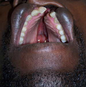
Postoperative infant feeding
Traditionally, breast pumping and specialized feeding (syringe or spoon) are used in the immediate postoperative period to minimize disturbance of the surgical site, but some surgeons advocate the use of more liberal feeding options. A randomized controlled trial of 232 infants aged 6 months and under with cleft palate or cleft lip showed a statistically significant improvement in weight at 6 weeks postsurgery when breastfeeding was used instead of spoon feeding, but no differences in growth outcomes were observed when rigid and flexible bottles were compared.[46]
Fetal surgical repair
Fetal surgical repair of cleft lip and palate requires surgical advances that minimize the risks to the mother and fetus. The embryonic midfacial structures organize from the 5 facial prominences at 4 to 5 weeks' gestation. Cleft lip and palate deformities occur due to an interruption of fusion of the maxillary and median nasal prominences and subsequent failure of mesoderm ingrowth. In the last few decades, fetal surgical repair of a variety of congenital malformations (e.g., congenital diaphragmatic hernia, twin-twin transfusion, cleft lip and palate) have been investigated. Early reports of fetal surgery show it heals with less inflammation and scarring.[74] Unfortunately, risks to the mother and fetus remain high, with a 25% fetal mortality rate. Maternal and fetal morbidity and mortality may be lower with fetal endoscopic surgical techniques. Both surgeons and parents would face difficult ethical dilemmas if fetal surgery, with its associated risks, were chosen as a method of treatment for improving lip scarring in an otherwise healthy fetus. A current consensus by the International Fetal Medicine and Surgery Society reserves fetal surgery for life-threatening conditions that have a poor prognosis when approached postnatally.[75]
OnabotulinumtoxinA (formerly known as botulinum toxin type A)
Repair of cleft lip defects requires meticulous attention to recreating the 3-dimensional characteristics of the lip and nasal deformity. The alignment of the orbicularis oris muscle is completed at cleft lip repair. However, its muscular contractions increase wound tension at the repair site. The success of the repair is influenced by wound tension during the healing phase. A widely separated cleft lip closed under too much tension may lead to wound breakdown with fistula formation or dehiscence, or heal with increased scarring. OnabotulinumtoxinA is hypothesized to improve the cosmetic outcome of cleft lip reconstruction by reducing wound tension, and its effectiveness has been studied using 3-dimensional videography to assess the decrease in lip movement postinjection.[76]
Use of this content is subject to our disclaimer