Images and videos
Images


Acute aspiration
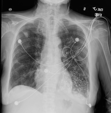
Barium aspiration. A barium swallow was conducted in a 53-year-old woman. Imaging revealed hyperdense airway-centered material in the left lower lobe consistent with barium aspiration bronchiolitis. A tracheoesophageal fistula was confirmed
From the collection of Dr Augustine Lee; used with permission of Mayo Foundation for Medical Education and Research, all rights reserved
See this image in context in the following section/s:

Acute aspiration
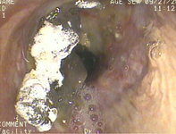
Bronchoscopy showing barium aspiration in a lung transplant patient in the right mainstem bronchus after a barium swallow study
From the collection of Dr Kamran Mahmood
See this image in context in the following section/s:
Acute aspiration
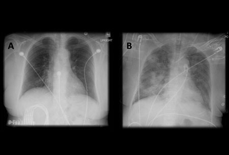
A. Portable upright chest x-ray before aspiration; B. Chest x-ray 1 hour after aspiration, showing bilateral diffuse alveolar infiltrates, worse at the bases on the right side
From the collection of Dr Henri Colt
See this image in context in the following section/s:
Acute aspiration
Lipoid pneumonia. A 77-year-old woman with dysphagia and achalasia following a stroke presented with recurrent lung infiltrates, including a persisting right middle lobe lesion. Hounsfield units (HU) measurement was -157, consistent with lipoid pneumonia. Comparative subcutaneous fat and aorta (blood/tissue) HU are shown
From the collection of Dr Augustine Lee, used with permission of Mayo Foundation for Medical Education and Research, all rights reserved
See this image in context in the following section/s:
Use of this content is subject to our disclaimer