One of the common settings in which pulmonary aspiration is known to take place is the perioperative period. Various risk factors including reduced consciousness, prolonged supine positioning, and illness acuity can predispose an individual to aspiration of gastric contents. In the US, reported incidence of perioperative pulmonary aspiration in the adult population ranges from 1 in 3216 (in 1993) to 1 in 7103 (in 2006).[5]Warner MA, Warner ME, Weber JG. Clinical significance of pulmonary aspiration during the perioperative period. Anesthesiology. 1993 Jan;78(1):56-62.
http://www.ncbi.nlm.nih.gov/pubmed/8424572?tool=bestpractice.com
[6]Sakai T, Planinsic RM, Quinlan JJ, et al. The incidence and outcome of perioperative pulmonary aspiration in a university hospital: a 4-year retrospective analysis. Anesth Analg. 2006 Oct;103(4):941-7.
https://journals.lww.com/anesthesia-analgesia/fulltext/2006/10000/the_incidence_and_outcome_of_perioperative.28.aspx
http://www.ncbi.nlm.nih.gov/pubmed/17000809?tool=bestpractice.com
Studies published in 1986 and 1993 reported that aspiration occurred in 1 in every 3000 cases of anesthesia and accounted for 10% to 30% of deaths associated with anesthesia.[2]Marik PE. Aspiration pneumonitis and aspiration pneumonia. N Engl J Med. 2001 Mar 1;344(9):665-71.
http://www.ncbi.nlm.nih.gov/pubmed/11228282?tool=bestpractice.com
[5]Warner MA, Warner ME, Weber JG. Clinical significance of pulmonary aspiration during the perioperative period. Anesthesiology. 1993 Jan;78(1):56-62.
http://www.ncbi.nlm.nih.gov/pubmed/8424572?tool=bestpractice.com
[7]Olsson GL, Hallen B, Hambraeus-Jonzon K. Aspiration during anaesthesia: a computer-aided study of 185,358 anaesthetics. Acta Anaesthesiol Scand. 1986 Jan;30(1):84-92.
http://www.ncbi.nlm.nih.gov/pubmed/3754372?tool=bestpractice.com
Although the incidence of aspiration is low, the extent of surgery has increased in people with comorbidities and the older population, and these patients are expected to have a higher incidence of aspiration.[8]Ng A, Smith G. Gastroesophageal reflux and aspiration of gastric contents in anesthetic practice. Anesth Analg. 2001 Aug;93(2):494-513.
http://www.anesthesia-analgesia.org/content/93/2/494.full
http://www.ncbi.nlm.nih.gov/pubmed/11473886?tool=bestpractice.com
In the pediatric population, one UK study published in 2013 notes an incidence of perioperative pulmonary aspiration of 1 in 4932 anesthetics in the elective setting and 1 in 4498 in the emergency setting.[9]Walker RW. Pulmonary aspiration in pediatric anesthetic practice in the UK: a prospective survey of specialist pediatric centers over a one-year period. Paediatr Anaesth. 2013 Aug;23(8):702-11.
http://www.ncbi.nlm.nih.gov/pubmed/23763657?tool=bestpractice.com
This suggests an improvement compared with two prior US landmark studies in children, which reported an incidence of 1 in 2632 aspirations during general anesthesia between 1985 and 1997, and 1 in 978 between 1988 and 1993.[10]Warner MA, Warner ME, Warner DO, et al. Perioperative pulmonary aspiration in infants and children. Anesthesiology. 1999 Jan;90(1):66-71.
http://www.ncbi.nlm.nih.gov/pubmed/9915314?tool=bestpractice.com
[11]Borland LM, Sereika SM, Woelfel SK, et al. Pulmonary aspiration in pediatric patients during general anesthesia: incidence and outcome. J Clin Anesth. 1998 Mar;10(2):95-102.
http://www.ncbi.nlm.nih.gov/pubmed/9524892?tool=bestpractice.com
Despite a low incidence, perioperative pulmonary aspiration carries a risk of morbidity and mortality. In US anesthesia malpractice claims data, 5% were attributed to aspiration, with a 57% mortality directly attributable to anesthesia.[12]Warner MA, Meyerhoff KL, Warner ME, et al. Pulmonary aspiration of gastric contents: a closed claims analysis. Anesthesiology. 2021 Aug 1;135(2):284-91.
https://journals.lww.com/anesthesiology/fulltext/2021/08000/pulmonary_aspiration_of_gastric_contents__a_closed.20.aspx
http://www.ncbi.nlm.nih.gov/pubmed/34019629?tool=bestpractice.com
Of those who aspirated, 61% had gastrointestinal obstruction or an acute abdomen.[12]Warner MA, Meyerhoff KL, Warner ME, et al. Pulmonary aspiration of gastric contents: a closed claims analysis. Anesthesiology. 2021 Aug 1;135(2):284-91.
https://journals.lww.com/anesthesiology/fulltext/2021/08000/pulmonary_aspiration_of_gastric_contents__a_closed.20.aspx
http://www.ncbi.nlm.nih.gov/pubmed/34019629?tool=bestpractice.com
The body of literature regarding acute aspiration outside of the perioperative time frame is smaller, and limited by the lack of specific diagnostic testing for aspiration when it is not witnessed. Between 5% and 15% of community-acquired pneumonia has been attributed to aspiration.[2]Marik PE. Aspiration pneumonitis and aspiration pneumonia. N Engl J Med. 2001 Mar 1;344(9):665-71.
http://www.ncbi.nlm.nih.gov/pubmed/11228282?tool=bestpractice.com
Pneumonia due to aspiration events is more frequent in patients ages >80 years (10%) compared with patients ages <80 years (5%).[13]Fernández-Sabé N, Carratalà J, Rosón B, et al. Community-acquired pneumonia in very elderly patients: causative organisms, clinical characteristics, and outcomes. Medicine (Baltimore). 2003 May;82(3):159-69.
https://journals.lww.com/md-journal/fulltext/2003/05000/community_acquired_pneumonia_in_very_elderly.2.aspx
http://www.ncbi.nlm.nih.gov/pubmed/12792302?tool=bestpractice.com
Predisposing factors for aspiration included dysphagia, and impaired consciousness and gag reflex.[13]Fernández-Sabé N, Carratalà J, Rosón B, et al. Community-acquired pneumonia in very elderly patients: causative organisms, clinical characteristics, and outcomes. Medicine (Baltimore). 2003 May;82(3):159-69.
https://journals.lww.com/md-journal/fulltext/2003/05000/community_acquired_pneumonia_in_very_elderly.2.aspx
http://www.ncbi.nlm.nih.gov/pubmed/12792302?tool=bestpractice.com
Another study noted that of a cohort of patients admitted with pneumonia ages >70 years, about 55% were found to have dysphagia and aspiration on testing with a water swallow test.[14]Cabre M, Serra-Prat M, Palomera E, et al. Prevalence and prognostic implications of dysphagia in elderly patients with pneumonia. Age Ageing. 2010 Jan;39(1):39-45.
http://www.ncbi.nlm.nih.gov/pubmed/19561160?tool=bestpractice.com
In the Lung Injury Prediction Score cohort, aspiration was the third leading cause of acute lung injury among at-risk patients admitted to the US hospitals.[15]Gajic O, Dabbagh O, Park PK, et al. Early identification of patients at risk of acute lung injury: evaluation of lung injury prediction score in a multicenter cohort study. Am J Respir Crit Care Med. 2011 Feb 15;183(4):462-70.
https://pmc.ncbi.nlm.nih.gov/articles/PMC3056224
http://www.ncbi.nlm.nih.gov/pubmed/20802164?tool=bestpractice.com
Dysphagia may cause "acute on chronic" aspirations. According to one systematic review from 2005, the incidence of dysphagia (swallowing difficulties) following acute stroke was reported to vary from 37% to 78% depending on the site of the stroke and screening tools used to identify dysphagia.[16]Intercollegiate Stroke Working Party. National clinical guideline for stroke for the UK and Ireland. May 2023 [internet publication].
https://www.strokeguideline.org
[17]Martino R, Foley N, Bhogal S, et al. Dysphagia after stroke: incidence, diagnosis, and pulmonary complications. Stroke. 2005 Dec;36(12):2756-63.
https://www.ahajournals.org/doi/10.1161/01.STR.0000190056.76543.eb
http://www.ncbi.nlm.nih.gov/pubmed/16269630?tool=bestpractice.com
A review from 2010 reported that up to 30% of older patients with dysphagia present with aspiration.[18]Rofes L, Arreola V, Almirall J, et al. Diagnosis and management of oropharyngeal dysphagia and its nutritional and respiratory complications in the elderly. Gastroenterol Res Pract. 2011:2011:818979.
https://onlinelibrary.wiley.com/doi/10.1155/2011/818979
http://www.ncbi.nlm.nih.gov/pubmed/20811545?tool=bestpractice.com
Among patients admitted to the hospital who are receiving enteral nutrition, the reported prevalence varies widely from 4.4% to nearly 90%, depending on how aspiration is defined (silent versus symptomatic), the method of diagnosis, the position of the feeding tube in the gastrointestinal tract (nasogastric or nasojejunal), and the type of feeding tube (nasogastric or gastrostomy).[19]Mullan H, Roubenoff RA, Roubenoff R. Risk of pulmonary aspiration among patients receiving enteral nutrition support. JPEN J Parenter Enteral Nutr. Mar-Apr 1992;16(2):160-4.
http://www.ncbi.nlm.nih.gov/pubmed/1556813?tool=bestpractice.com
[20]Metheny NA, Clouse RE, Chang YH, et al. Tracheobronchial aspiration of gastric contents in critically ill tube-fed patients: frequency, outcomes, and risk factors. Crit Care Med. 2006 Apr;34(4):1007-15.
http://www.ncbi.nlm.nih.gov/pmc/articles/PMC2396145
http://www.ncbi.nlm.nih.gov/pubmed/16484901?tool=bestpractice.com
One meta-analysis conducted in 2019 including 41 studies and involving 3248 participants suggests there is a lower incidence of pulmonary aspiration with postpyloric feeding tube positioning compared with gastric positioning.[21]Liu Y, Wang Y, Zhang B, et al. Gastric-tube versus post-pyloric feeding in critical patients: a systematic review and meta-analysis of pulmonary aspiration- and nutrition-related outcomes. Eur J Clin Nutr. 2021 Sep;75(9):1337-48.
https://www.nature.com/articles/s41430-021-00860-2
http://www.ncbi.nlm.nih.gov/pubmed/33536570?tool=bestpractice.com
Clinically evident aspiration, however, is rare and is seen in <1% of patients given enteral nutrition.[20]Metheny NA, Clouse RE, Chang YH, et al. Tracheobronchial aspiration of gastric contents in critically ill tube-fed patients: frequency, outcomes, and risk factors. Crit Care Med. 2006 Apr;34(4):1007-15.
http://www.ncbi.nlm.nih.gov/pmc/articles/PMC2396145
http://www.ncbi.nlm.nih.gov/pubmed/16484901?tool=bestpractice.com
In the setting of severe trauma, the incidence of gross aspiration can be as high as 38%.[22]Lockey DJ, Coats T, Parr MJ. Aspiration in severe trauma: a prospective study. Anaesthesia. 1999 Nov;54(11):1097-8.
https://onlinelibrary.wiley.com/doi/full/10.1046/j.1365-2044.1999.00754.x?sid=nlm%3Apubmed
http://www.ncbi.nlm.nih.gov/pubmed/10540100?tool=bestpractice.com
Accidental aspiration of barium contrast medium during radiologic investigations is rare but can occur in up to 8% of children with GERD.[23]Fung KP, Seagram G, Pasieka J, et al. Investigation and outcome of 121 infants and children requiring Nissen fundoplication for the management of gastroesophageal reflux. Clin Invest Med. 1990 Oct;13(5):237-46.
http://www.ncbi.nlm.nih.gov/pubmed/2276217?tool=bestpractice.com
The severity of pulmonary damage depends on the density of the suspension, with high-density barium sulfate causing the most damage and being potentially fatal, especially in older patients.[4]Gray C, Sivaloganathan S, Simpkins KC. Aspiration of high-density barium contrast medium causing acute pulmonary inflammation: report of two fatal cases in elderly women with disordered swallowing. 1989 Jul;40(4):397-400.
http://www.ncbi.nlm.nih.gov/pubmed/2758750?tool=bestpractice.com
[24]Tamm I, Kortsik C. Severe barium sulfate aspiration into the lung: clinical presentation, prognosis and therapy. Respiration. 1999;66(1):81-4.
http://www.ncbi.nlm.nih.gov/pubmed/9973698?tool=bestpractice.com
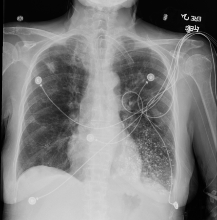
[25]Ansell G. Alimentary tract. In: Ansell G, Bettmann MA, Kaufman JA, et al, eds. Complications in diagnostic imaging and interventional radiology. 2nd ed. Oxford, UK: Blackwell Scientific; 1987:218-9.[Figure caption and citation for the preceding image starts]: Bronchoscopy showing barium aspiration in a lung transplant patient in the right mainstem bronchus after a barium swallow studyFrom the collection of Dr Kamran Mahmood [Citation ends]. [Figure caption and citation for the preceding image starts]: Barium aspiration. A barium swallow was conducted in a 53-year-old woman. Imaging revealed hyperdense airway-centered material in the left lower lobe consistent with barium aspiration bronchiolitis. A tracheoesophageal fistula was confirmedFrom the collection of Dr Augustine Lee; used with permission of Mayo Foundation for Medical Education and Research, all rights reserved [Citation ends].