Images and videos
Images


Cerebral aneurysm
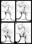
Progressive angiography images of a small dissecting aneurysm of the distal basilar artery after a subarachnoid and intraventricular hemorrhage on day 3 (A), day 23 (B), and day 30 (C), and 6 months after stent-assisted coiling (D). Arrows indicate proximal and distal stent markers
From: Peluso JP, van Rooij WJ, Sluzewski M. BMJ Case Reports 2009; doi:10.1136/bcr.2007.121533. Used with permission
See this image in context in the following section/s:

Cerebral aneurysm
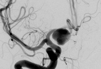
Cerebral angiogram showing aneurysm
From the personal collection of Dr M. Chen, Columbia College of Physicians and Surgeons
See this image in context in the following section/s:
Cerebral aneurysm
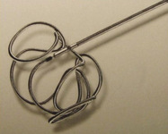
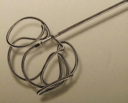
Example of a coil used to treat cerebral aneurysms
From: Sellar M. Practical Neurology. 2005;5:28-37. Used with permission
See this image in context in the following section/s:
Cerebral aneurysm
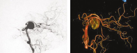
Comparison of 2-dimensional catheter angiography (left) with 3-dimensional catheter angiography (right) showing a basilar tip aneurysm
From: Sellar M. Practical Neurology. 2005;5:28-37. Used with permission
See this image in context in the following section/s:

Cerebral aneurysm
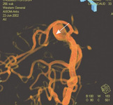
Three-dimensional catheter angiogram showing a basilar tip aneurysm
From: Sellar M. Practical Neurology. 2005;5:28-37. Used with permission
See this image in context in the following section/s:
Use of this content is subject to our disclaimer