Images and videos
Images
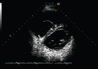


Pulmonary regurgitation
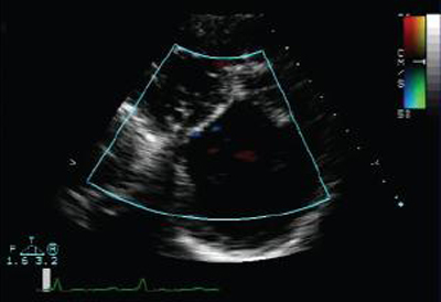
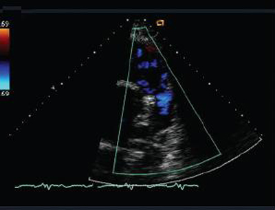
Short axis echocardiographic view in a patient with pulmonary regurgitation following repair of tetralogy of Fallot. The patient has a restrictive right ventricle, and short axis echocardiographic view reveals a small right ventricle
From: Chaturvedi RR, Redington AN. Heart. 2007 Jul;93(7):880-9; used with permission
See this image in context in the following section/s:

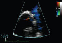
Pulmonary regurgitation
Short axis echocardiographic view in a patient with pulmonary regurgitation following repair of tetralogy of Fallot. The patient has a restrictive right ventricle, and short axis echocardiographic view reveals that the right ventricle is large
From: Chaturvedi RR, Redington AN. Heart. 2007 Jul;93(7):880-9; used with permission
See this image in context in the following section/s:

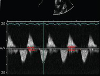
Pulmonary regurgitation
Doppler in a patient with pulmonary regurgitation following repair of tetralogy of Fallot. The patient has a restrictive right ventricle, and Doppler shows evidence of restriction with an antegrade "A" wave in the pulmonary artery
From: Chaturvedi RR, Redington AN. Heart. 2007 Jul;93(7):880-9; used with permission
See this image in context in the following section/s:
Pulmonary regurgitation
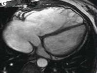
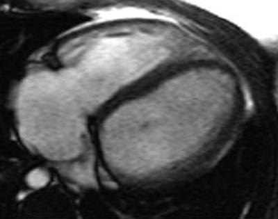
Magnetic resonance imaging (MRI) in a patient with pulmonary regurgitation following repair of tetralogy of Fallot. The patient has a restrictive right ventricle, and MRI shows decreased right ventricular volume
From: Chaturvedi RR, Redington AN. Heart. 2007 Jul;93(7):880-9; used with permission
See this image in context in the following section/s:
Pulmonary regurgitation
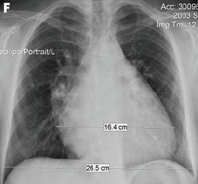
Chest x-ray in a patient with pulmonary regurgitation following repair of tetralogy of Fallot. The patient has a restrictive right ventricle and the heart is small
From: Chaturvedi RR, Redington AN. Heart. 2007 Jul;93(7):880-9; used with permission
See this image in context in the following section/s:
Pulmonary regurgitation
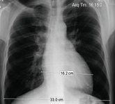
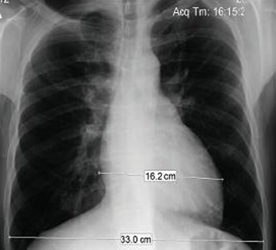
Chest x-ray in a patient with pulmonary regurgitation following repair of tetralogy of Fallot. The patient has a nonrestrictive right ventricle and the heart is large
From: Chaturvedi RR, Redington AN. Heart. 2007 Jul;93(7):880-9; used with permission
See this image in context in the following section/s:
Pulmonary regurgitation
Doppler echocardiogram in a patient with pulmonary regurgitation following repair of tetralogy of Fallot, revealing a nonobstructed right ventricular outflow tract. The patient has a nonrestrictive right ventricle
From: Chaturvedi RR, Redington AN. Heart. 2007 Jul;93(7):880-9; used with permission
See this image in context in the following section/s:

Pulmonary regurgitation
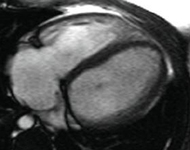
Magnetic resonance imaging (MRI) in a patient with pulmonary regurgitation following repair of tetralogy of Fallot. The patient has a nonrestrictive right ventricle, and MRI shows that the right ventricle is dilated
From: Chaturvedi RR, Redington AN. Heart. 2007 Jul;93(7):880-9; used with permission
See this image in context in the following section/s:

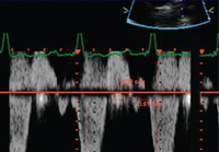
Pulmonary regurgitation
Doppler in a patient with pulmonary regurgitation following repair of tetralogy of Fallot. The patient has a nonrestrictive right ventricle, and an ‘‘A’’ wave is not seen in the pulmonary artery Doppler spectrum
From: Chaturvedi RR, Redington AN. Heart. 2007 Jul;93(7):880-9; used with permission
See this image in context in the following section/s:

Pulmonary regurgitation
Doppler echocardiogram in a patient with pulmonary regurgitation following repair of tetralogy of Fallot, revealing a non-obstructed right ventricular outflow tract. The patient has a restrictive right ventricle
From: Chaturvedi RR, Redington AN. Heart. 2007 Jul;93(7):880-9; used with permission
See this image in context in the following section/s:
Use of this content is subject to our disclaimer