The treatment of long bone fractures is determined by the specific fracture type, nature, and severity. If the patient is stable, a splint should be applied to the affected extremity to provide immobilization and protection.
Adequate analgesia should be provided, and x-rays should be obtained while waiting for the orthopedic surgeon. If fracture displacement and deformity lead to neurovascular compromise or inability to splint or transport the patient, gentle in-line traction may be attempted to reduce the fracture. Venous thromboembolism prophylaxis should be considered according to current guidance.[74]Falck-Ytter Y, Francis CW, Johanson NA, et al ; American College of Chest Physicians. Prevention of VTE in orthopedic surgery patients: antithrombotic therapy and prevention of thrombosis, 9th ed: American College of Chest Physicians evidence-based clinical practice guidelines. Chest. 2012 Feb;141(2 Suppl):e278S-e325S.
https://journal.publications.chestnet.org/article.aspx?articleid=1159591
http://www.ncbi.nlm.nih.gov/pubmed/22315265?tool=bestpractice.com
[75]ICM-VTE General Delegates. Recommendations from the ICM-VTE: General. J Bone Joint Surg Am. 2022 Mar 16;104(suppl 1):4-162.
https://www.doi.org/10.2106/JBJS.21.01531
http://www.ncbi.nlm.nih.gov/pubmed/35315607?tool=bestpractice.com
[76]Douketis JD, Spyropoulos AC, Murad MH, et al. Perioperative management of antithrombotic therapy: an American College of Chest Physicians clinical practice guideline. Chest. 2022 Nov;162(5):e207-43.
https://www.doi.org/10.1016/j.chest.2022.07.025
http://www.ncbi.nlm.nih.gov/pubmed/35964704?tool=bestpractice.com
The American Academy of Orthopaedic Surgeons (AAOS) recommends that patients with open fractures are brought to the operating room for debridement and irrigation as soon as possible, and ideally within 24 hours post injury.[77]American Academy of Orthopaedic Surgeons. Prevention of surgical site infections after major extremity trauma. Evidence-based clinical practice guideline. Mar 2022 [internet publication].
https://www.aaos.org/globalassets/quality-and-practice-resources/dod/ssitrauma/ssitraumacpg.pdf
[78]American Academy of Orthopaedic Surgeons. Appropriate use criteria: distal radius fractures: treatment. 2013 [internet publication].
https://www.aaos.org/globalassets/quality-and-practice-resources/distal-radius/drf_auc.pdf
After closed fracture fixation, negative pressure wound therapy may mitigate the risk of revision surgery or surgical site infections. However, after open fracture fixation, negative pressure wound therapy does not appear to offer an advantage when compared with sealed dressings as it does not decrease wound complications or amputations.[77]American Academy of Orthopaedic Surgeons. Prevention of surgical site infections after major extremity trauma. Evidence-based clinical practice guideline. Mar 2022 [internet publication].
https://www.aaos.org/globalassets/quality-and-practice-resources/dod/ssitrauma/ssitraumacpg.pdf
[78]American Academy of Orthopaedic Surgeons. Appropriate use criteria: distal radius fractures: treatment. 2013 [internet publication].
https://www.aaos.org/globalassets/quality-and-practice-resources/distal-radius/drf_auc.pdf
Silver-coated dressings are not recommended as they do not improve outcomes or decrease pin-site infections.[77]American Academy of Orthopaedic Surgeons. Prevention of surgical site infections after major extremity trauma. Evidence-based clinical practice guideline. Mar 2022 [internet publication].
https://www.aaos.org/globalassets/quality-and-practice-resources/dod/ssitrauma/ssitraumacpg.pdf
[78]American Academy of Orthopaedic Surgeons. Appropriate use criteria: distal radius fractures: treatment. 2013 [internet publication].
https://www.aaos.org/globalassets/quality-and-practice-resources/distal-radius/drf_auc.pdf
[Figure caption and citation for the preceding image starts]: Recommended immobilization techniques for long bone fracturesCreated by the BMJ Evidence Centre [Citation ends].
High-energy trauma
Most acute long bone shaft (diaphyseal) fractures are caused by high-energy trauma and are often associated with other, potentially life-threatening injuries. In these situations, a complete head-to-toe exam must be performed, with institution of Advanced Trauma Life Support (ATLS)/Advanced Cardiac Life Support (ACLS) methods to ensure hemodynamic stability and prevent further injury. The appropriate local or national resuscitation and management protocols should be followed. For patients with severe acute hemorrhage, antifibrinolytics (e.g. tranexamic acid) should be considered, because these agents have been shown to increase survival.[79]Ker K, Roberts I, Shakur H, et al. Antifibrinolytic drugs for acute traumatic injury. Cochrane Database Syst Rev. 2015 May 9;(5):CD004896.
https://www.doi.org/10.1002/14651858.CD004896.pub4
http://www.ncbi.nlm.nih.gov/pubmed/25956410?tool=bestpractice.com
[80]CRASH-2 collaborators., Roberts I, Shakur H, et al. The importance of early treatment with tranexamic acid in bleeding trauma patients: an exploratory analysis of the CRASH-2 randomised controlled trial. Lancet. 2011 Mar 26;377(9771):1096-101, 1101.e1-2.
https://www.doi.org/10.1016/S0140-6736(11)60278-X
http://www.ncbi.nlm.nih.gov/pubmed/21439633?tool=bestpractice.com
Delay in administration reduces their benefit; delays in administration of tranexamic acid were associated with reduced survival in a meta-analysis of data from patients with traumatic bleeding or postpartum hemorrhage (survival benefit decreasing by about 10% for every 15 minutes of treatment delay until 3 hours, after which there was no benefit).[81]Gayet-Ageron A, Prieto-Merino D, Ker K, et al. Effect of treatment delay on the effectiveness and safety of antifibrinolytics in acute severe haemorrhage: a meta-analysis of individual patient-level data from 40 138 bleeding patients. Lancet. 2018 Jan 13;391(10116):125-32.
https://www.doi.org/10.1016/S0140-6736(17)32455-8
http://www.ncbi.nlm.nih.gov/pubmed/29126600?tool=bestpractice.com
Hypotension, hypovolemic shock, compartment syndrome, fat embolism syndrome, and further hemorrhage may ensue, so rapid, thorough evaluation and serial exams are of paramount importance. Urgent or emergent orthopedic consultation is required, as operative treatment is the preferred approach for most of these injuries.
Distal humeral shaft fractures
If the patient is stable and has an isolated, nondisplaced humeral shaft fracture, treatment may consist of splint immobilization, elevation, ice, and analgesia. Consultation with an orthopedic surgeon is recommended. Most displaced humeral shaft fractures heal well with nonoperative management (i.e., coaptation [or sugar-tong] splinting). Operative intervention is required if fracture alignment is unacceptable after closed reduction.
Midshaft humeral fracture
Closed midshaft humeral fractures tend to heal fairly well with nonoperative management. A transverse fracture may be treated initially with a coaptation (or sugar-tong) splint and sling, and subsequently with functional bracing. One clinical trial suggested that surgical intervention (i.e., compression plating) for midshaft humeral fractures may have a lower rate of nonunion and malunion than nonoperative treatment.[82]Denard A Jr, Richards JE, Obremskey WT, et al. Outcome of nonoperative vs operative treatment of humeral shaft fractures: a retrospective study of 213 patients. Orthopedics. 2010 Aug 11;33(8).
http://www.ncbi.nlm.nih.gov/pubmed/20704103?tool=bestpractice.com
However, the optimal treatment of these fractures is still not clear, based on the relative lack of high-quality evidence.[83]Gosler MW, Testroote M, Morrenhof JW, et al. Surgical versus non-surgical interventions for treating humeral shaft fractures in adults. Cochrane Database Syst Rev. 2012 Jan 18;1:CD008832.
https://onlinelibrary.wiley.com/doi/10.1002/14651858.CD008832.pub2/full
http://www.ncbi.nlm.nih.gov/pubmed/22258990?tool=bestpractice.com
Physical therapy with early mobilization is considered important to restore function and minimize the chance of adhesive capsulitis.
Fractures in which adequate positioning cannot be achieved/maintained, or which are grossly unstable, should be treated operatively. It is unclear whether dynamic compression plating is superior to intramedullary nailing, although plating may reduce the risk of impingement.[84]Ma J, Xing D, Ma X, et al. Intramedullary Nail versus Dynamic Compression Plate Fixation in Treating Humeral Shaft Fractures: Grading the Evidence through a Meta-Analysis. PLoS One. 2013 Dec 16;8(12):e82075.
https://www.ncbi.nlm.nih.gov/pmc/articles/PMC3864910
http://www.ncbi.nlm.nih.gov/pubmed/24358141?tool=bestpractice.com
Proximal humeral shaft fractures
Management of proximal humeral shaft fractures depends on the Neer classification: 1-part fractures generally do well with conservative management; analgesia, ice, and immobilization in a sling are generally followed by institution of early range of motion exercises; and 2-, 3-, and 4-part fractures require orthopedic surgery evaluation.[4]Neer CS 2nd. Displaced proximal humeral fractures: part I. Classification and evaluation. J Bone Joint Surg Am. 1970 Sep;52(6):1077-89.
https://jbjs.org/article.aspx?articleid=15414
In the Neer system, the fracture is classified by involvement and displacement of the following 4 structural segments:
Greater tuberosity
Lesser tuberosity
Humeral head
Humeral shaft.
Although many fracture lines may be seen, if no displacement is present (defined as <1 cm of separation and <45° of angulation), it is considered a 1-part fracture. If only 1 segment is displaced, a 2-part fracture is present. If 2 segments are displaced, a 3-part fracture is present. If all 4 segments are displaced, a 4-part fracture is present.
Intra-articular fractures, associated dislocation, suspected rotator cuff tear, or fractures of the surgical neck are indications for urgent orthopedic evaluation.[85]Handoll HH, Elliott J, Thillemann TM, et al. Interventions for treating proximal humeral fractures in adults. Cochrane Database Syst Rev. 2022 Jun 21;6(6):CD000434.
https://www.cochranelibrary.com/cdsr/doi/10.1002/14651858.CD000434.pub5/full
http://www.ncbi.nlm.nih.gov/pubmed/35727196?tool=bestpractice.com
Emergency orthopedic and vascular surgery consultation is also required for any suspected neurovascular injury.
One Cochrane review of 10 trials (717 participants) concluded there is high- or moderate-certainty evidence that, compared with nonsurgical treatment, surgery does not result in a better outcome at 1 and 2 years after injury for people aged 60 years and over with displaced proximal humeral fractures. A surgical approach may increase the need for subsequent surgery.[85]Handoll HH, Elliott J, Thillemann TM, et al. Interventions for treating proximal humeral fractures in adults. Cochrane Database Syst Rev. 2022 Jun 21;6(6):CD000434.
https://www.cochranelibrary.com/cdsr/doi/10.1002/14651858.CD000434.pub5/full
http://www.ncbi.nlm.nih.gov/pubmed/35727196?tool=bestpractice.com
[  ]
How does surgical intervention compare with non‐surgical treatment for treating proximal humeral fractures in adults?/cca.html?targetUrl=https://www.cochranelibrary.com/cca/doi/10.1002/cca.4137/fullShow me the answer There is insufficient evidence from randomized controlled trials to compare surgical versus nonsurgical approaches for people aged under 60 years, high-energy trauma, two-part tuberosity fractures, or less common fractures, such as fracture dislocations and articular surface fractures.[85]Handoll HH, Elliott J, Thillemann TM, et al. Interventions for treating proximal humeral fractures in adults. Cochrane Database Syst Rev. 2022 Jun 21;6(6):CD000434.
https://www.cochranelibrary.com/cdsr/doi/10.1002/14651858.CD000434.pub5/full
http://www.ncbi.nlm.nih.gov/pubmed/35727196?tool=bestpractice.com
Close collaboration with an experienced orthopedic surgeon is recommended, and the choice between surgical versus nonsurgical approaches should be individualized.
]
How does surgical intervention compare with non‐surgical treatment for treating proximal humeral fractures in adults?/cca.html?targetUrl=https://www.cochranelibrary.com/cca/doi/10.1002/cca.4137/fullShow me the answer There is insufficient evidence from randomized controlled trials to compare surgical versus nonsurgical approaches for people aged under 60 years, high-energy trauma, two-part tuberosity fractures, or less common fractures, such as fracture dislocations and articular surface fractures.[85]Handoll HH, Elliott J, Thillemann TM, et al. Interventions for treating proximal humeral fractures in adults. Cochrane Database Syst Rev. 2022 Jun 21;6(6):CD000434.
https://www.cochranelibrary.com/cdsr/doi/10.1002/14651858.CD000434.pub5/full
http://www.ncbi.nlm.nih.gov/pubmed/35727196?tool=bestpractice.com
Close collaboration with an experienced orthopedic surgeon is recommended, and the choice between surgical versus nonsurgical approaches should be individualized.
Radial and ulnar shaft fractures
Initial treatment of radial fractures includes placement of a splint and urgent orthopedic referral. Optimal treatment involves open reduction and internal fixation (ORIF) of the fracture when the wound is determined to be clean, along with stabilization of the distal radioulnar joint in cases of a Galeazzi fracture.[77]American Academy of Orthopaedic Surgeons. Prevention of surgical site infections after major extremity trauma. Evidence-based clinical practice guideline. Mar 2022 [internet publication].
https://www.aaos.org/globalassets/quality-and-practice-resources/dod/ssitrauma/ssitraumacpg.pdf
[86]Macintyre NR, Ilyas AM, Jupiter JB. Treatment of forearm fractures. Acta Chir Orthop Traumatol Cech. 2009 Feb;76(1):7-14.
http://www.ncbi.nlm.nih.gov/pubmed/19268042?tool=bestpractice.com
[87]American Academy of Orthopaedic Surgeons. Appropriate use criteria: prevention of surgical site infection after high energy extremity trauma. 2022 [internet publication].
https://www.orthoguidelines.org/go/auc/auc.cfm?auc_id=225052
Associated injury of the triangular fibrocartilage complex may need to be addressed surgically as well.
Treatment of nondisplaced fractures involves splinting and then conversion to a functional forearm brace, although no clearly superior approach has been demonstrated.[85]Handoll HH, Elliott J, Thillemann TM, et al. Interventions for treating proximal humeral fractures in adults. Cochrane Database Syst Rev. 2022 Jun 21;6(6):CD000434.
https://www.cochranelibrary.com/cdsr/doi/10.1002/14651858.CD000434.pub5/full
http://www.ncbi.nlm.nih.gov/pubmed/35727196?tool=bestpractice.com
Displaced or comminuted fractures require orthopedic referral for ORIF.
Fracture involving the proximal third of the ulna plus associated dislocation of the radial head (Monteggia fracture) requires urgent orthopedic consultation for ORIF. Long-term complications include heterotopic ossification at the elbow.[88]Eathiraju S, Mudgal CS, Jupiter JB. Monteggia fracture-dislocations. Hand Clin. 2007 May;23(2):165-77, v.
http://www.ncbi.nlm.nih.gov/pubmed/17548008?tool=bestpractice.com
A sugar-tong splint is recommended for initial immobilization of most forearm fractures; however, a double sugar-tong splint would be used in Monteggia fractures (or other elbow fractures).[Figure caption and citation for the preceding image starts]: Sugar-tong splintAuthor (Philip Cohen) [Citation ends].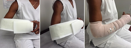 [Figure caption and citation for the preceding image starts]: Double sugar-tong splintAuthor (Philip Cohen) [Citation ends].
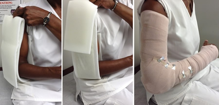
Stress fractures of the upper limb
These are generally treated with relative rest, analgesia, and a physical rehabilitation program.
Femoral shaft fractures
Acute treatment involves immediate orthopedic consultation while a thorough trauma evaluation and ATLS/ACLS measures are instituted. A traction splint can provide immobilization and pain relief, but in patients with multiple injuries or open fracture, such splinting may be impractical.[89]Wood SP, Vrahas M, Wedel SK. Femur fracture immobilization with traction splints in multisystem trauma patients. Prehosp Emerg Care. 2003 Apr-Jun;7(2):241-3.
http://www.ncbi.nlm.nih.gov/pubmed/12710786?tool=bestpractice.com
Adequate analgesia should also be given. In patients with a femoral shaft fracture who are awaiting surgical intervention, a femoral nerve block may provide superior anesthesia to a fascia iliaca compartment block, or to isolated parenteral morphine.[90]Newman B, McCarthy L, Thomas PW, et al. A comparison of pre-operative nerve stimulator-guided femoral nerve block and fascia iliaca compartment block in patients with a femoral neck fracture. Anaesthesia. 2013 Sep;68(9):899-903.
http://www.ncbi.nlm.nih.gov/pubmed/23789738?tool=bestpractice.com
[91]Beaudoin FL, Haran JP, Liebmann O. A comparison of ultrasound-guided three-in-one femoral nerve block versus parenteral opioids alone for analgesia in emergency department patients with hip fractures: a randomized controlled trial. Acad Emerg Med. 2013 Jun;20(6):584-91.
http://www.ncbi.nlm.nih.gov/pubmed/23758305?tool=bestpractice.com
However, for adult femoral shaft fractures, there is very little evidence to inform the use of this technique. One concern has been that a femoral nerve block might mask the symptoms of a developing compartment syndrome. One randomized trial compared intravenous fentanyl with femoral nerve block prior to spinal anesthesia for surgical intervention for femoral shaft fracture. Femoral nerve block was found to have better patient acceptance, to be associated with lower pain ratings, and to allow better positioning for spinal anesthesia.[92]Sia S, Pelusio F, Barbagli R, et al. Analgesia before performing a spinal block in the sitting position in patients with femoral shaft fracture: a comparison between femoral nerve block and intravenous fentanyl. Anesth Analg. 2004 Oct;99(4):1221-4.
http://www.ncbi.nlm.nih.gov/pubmed/15385380?tool=bestpractice.com
One review found no evidence to suggest that femoral nerve block delayed the diagnosis of compartment syndrome.[93]Karagiannis G, Hardern R. Best evidence topic report. No evidence found that a femoral nerve block in cases of femoral shaft fractures can delay the diagnosis of compartment syndrome of the thigh. Emerg Med J. 2005 Nov;22(11):814.
https://www.ncbi.nlm.nih.gov/pmc/articles/PMC1726592/pdf/v022p00814.pdf
http://www.ncbi.nlm.nih.gov/pubmed/16244347?tool=bestpractice.com
Intramedullary nailing is the preferred treatment for most femoral shaft fractures. Anterograde nailing is generally used, but in certain situations (distal femoral fracture, obese or pregnant patients, or patients who have undergone ipsilateral total hip arthroplasty), retrograde nailing may be useful.[94]Neubauer T, Ritter E, Potschka T, et al. Retrograde nailing of femoral fractures. Acta Chir Orthop Traumatol Cech. 2008 Jun;75(3):158-66.
http://www.ncbi.nlm.nih.gov/pubmed/18601812?tool=bestpractice.com
Tibial and fibular shaft fractures
Displaced, comminuted, and open fractures require immediate orthopedic consultation after initial immobilization with a splint and adequate analgesia. The treatment for shaft fractures is intramedullary nailing. More proximal and more distal fractures require ORIF.
A closed fracture that is nondisplaced and not comminuted can initially be treated with nonweight bearing and splint immobilization, with subsequent conversion to a long leg cast, although functional bracing for truly nondisplaced tibial shaft fractures is commonly used.[95]Bara T, Sibinski M, Synder M. Own clinical experience with functional bracing for treatment of pseudarthrosis and delayed union of the tibia. Ortop Traumatol Rehabil. 2007 May-Jun;9(3):259-63.
http://www.ncbi.nlm.nih.gov/pubmed/17721423?tool=bestpractice.com
[96]Sarmiento A, Latta LL. 450 closed fractures of the distal third of the tibia treated with a functional brace. Clin Orthop Relat Res. 2004 Nov;(428):261-71.
http://www.ncbi.nlm.nih.gov/pubmed/15534552?tool=bestpractice.com
An isolated fibular fracture usually heals well with conservative care (initial nonweight bearing, followed by transition to long leg walking cast, cast boot, or compression brace). [Figure caption and citation for the preceding image starts]: Posterior leg splintAuthor (Philip Cohen) [Citation ends].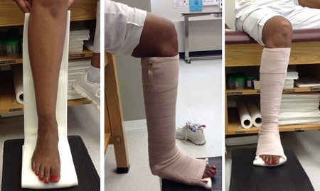
Stress fractures of the lower limb
Femoral stress fractures generally heal well with pain-free nonimpact cross-training and addressing underlying risk factors. However, a patient suspected of having a femoral neck stress fracture should be made nonweight bearing immediately and referred for urgent x-rays of the hip and proximal femur. If the films reveal a tension side fracture, a frank fracture line, or a displaced fracture, urgent orthopedic referral is needed for consideration of operative intervention. If the films reveal sclerosis at the compression side, an experienced provider may feel comfortable following the patient with serial radiographs and having them progress to partial then full weight bearing as tolerated. If the films are negative (common early on in the evolution of the fracture), a triple-phase bone scan (TPBS) or MRI can be used to detect the fracture. If the x-rays are negative but the TPBS is positive, conservative management by an experienced provider is reasonable. Full return to impact activity can take several months.[97]Kaeding CC, Yu JR, Wright R, et al. Management and return to play of stress fractures. Clin J Sport Med. 2005 Nov;15(6):442-7.
http://www.ncbi.nlm.nih.gov/pubmed/16278549?tool=bestpractice.com
Posteromedial tibial stress fractures are usually treated with modified weight bearing as tolerated and cessation of impact activity. Pain-free nonimpact cross-training (deep-water pool running, exercise biking, etc.) can be used to maintain fitness. Some studies have shown that the use of a pneumatic compression brace may allow the fracture to heal faster so that the patient can return to impact activity sooner.[98]Dickson TB Jr, Kichline PD. Functional management of stress fractures in female athletes using a pneumatic leg brace. Am J Sports Med. 1987 Jan-Feb;15(1):86-9.
http://www.ncbi.nlm.nih.gov/pubmed/3812866?tool=bestpractice.com
[99]Swenson EJ Jr, DeHaven KE, Sebastianelli WJ, et al. The effect of a pneumatic leg brace on return to play in athletes with tibial stress fractures. Am J Sports Med. 1997 May-Jun;25(3):322-8.
http://www.ncbi.nlm.nih.gov/pubmed/9167811?tool=bestpractice.com
Addressing biomechanical issues (e.g., over-pronation), insuring proper footwear, and preventing over-training are important to prevent recurrences.
Stress fractures of the fibula are uncommon but typically occur in runners and ballet dancers. Initial x-ray findings may be negative, but TPBS or MRI can demonstrate the fracture earlier. Treatment includes modified activity and transition to weight bearing as tolerated. As with other stress fractures, addressing training errors and other potentially modifiable risk factors is important, as is assessing for the possibility of eating disorders and related conditions.[100]Monteleone GP Jr. Stress fractures in the athlete. Orthop Clin North Am. 1995 Jul;26(3):423-32.
http://www.ncbi.nlm.nih.gov/pubmed/7609957?tool=bestpractice.com
Open fractures
The AAOS recommends that patients with open fractures are brought to the operating room for debridement and irrigation with saline as soon as reasonable, and ideally within 24 hours post injury.[77]American Academy of Orthopaedic Surgeons. Prevention of surgical site infections after major extremity trauma. Evidence-based clinical practice guideline. Mar 2022 [internet publication].
https://www.aaos.org/globalassets/quality-and-practice-resources/dod/ssitrauma/ssitraumacpg.pdf
[87]American Academy of Orthopaedic Surgeons. Appropriate use criteria: prevention of surgical site infection after high energy extremity trauma. 2022 [internet publication].
https://www.orthoguidelines.org/go/auc/auc.cfm?auc_id=225052
This has been shown to help prevent infections.[101]Tripuraneni K, Ganga S, Quinn R, et al. The effect of time delay to surgical debridement of open tibia shaft fractures on infection rate. Orthopedics. 2008 Dec;31(12).
https://www.orthosupersite.com/view.asp?rID=32925
http://www.ncbi.nlm.nih.gov/pubmed/19226070?tool=bestpractice.com
In addition, a plastic surgery consultation may be sought as open fractures may require soft tissue coverage and this should be done within the first 5 days.
The decision to perform ORIF or external fixation will depend upon the exact nature and severity of the injury, as well as the overall status of the patient. Temporizing external fixation remains a viable option for the treatment of open fractures in major extremity trauma.[77]American Academy of Orthopaedic Surgeons. Prevention of surgical site infections after major extremity trauma. Evidence-based clinical practice guideline. Mar 2022 [internet publication].
https://www.aaos.org/globalassets/quality-and-practice-resources/dod/ssitrauma/ssitraumacpg.pdf
[87]American Academy of Orthopaedic Surgeons. Appropriate use criteria: prevention of surgical site infection after high energy extremity trauma. 2022 [internet publication].
https://www.orthoguidelines.org/go/auc/auc.cfm?auc_id=225052
Definitive fixation of fractures at initial debridement and primary closure of wounds in selected patients may be considered when appropriate.[77]American Academy of Orthopaedic Surgeons. Prevention of surgical site infections after major extremity trauma. Evidence-based clinical practice guideline. Mar 2022 [internet publication].
https://www.aaos.org/globalassets/quality-and-practice-resources/dod/ssitrauma/ssitraumacpg.pdf
[87]American Academy of Orthopaedic Surgeons. Appropriate use criteria: prevention of surgical site infection after high energy extremity trauma. 2022 [internet publication].
https://www.orthoguidelines.org/go/auc/auc.cfm?auc_id=225052
For external fixation, fine wire fixators (Ilizarov frames) can be used.
Early delivery of antibiotics is suggested to lower the risk of deep infection in the setting of open fracture in major extremity trauma.[77]American Academy of Orthopaedic Surgeons. Prevention of surgical site infections after major extremity trauma. Evidence-based clinical practice guideline. Mar 2022 [internet publication].
https://www.aaos.org/globalassets/quality-and-practice-resources/dod/ssitrauma/ssitraumacpg.pdf
[87]American Academy of Orthopaedic Surgeons. Appropriate use criteria: prevention of surgical site infection after high energy extremity trauma. 2022 [internet publication].
https://www.orthoguidelines.org/go/auc/auc.cfm?auc_id=225052
In patients with major extremity trauma undergoing surgery, the AAOS strongly recommends that antibiotic prophylaxis with systemic cefazolin or clindamycin be administered, except for type III (and possibly type II) open fractures, for which additional gram-negative coverage (e.g., piperacillin/tazobactam) is preferred.[77]American Academy of Orthopaedic Surgeons. Prevention of surgical site infections after major extremity trauma. Evidence-based clinical practice guideline. Mar 2022 [internet publication].
https://www.aaos.org/globalassets/quality-and-practice-resources/dod/ssitrauma/ssitraumacpg.pdf
[87]American Academy of Orthopaedic Surgeons. Appropriate use criteria: prevention of surgical site infection after high energy extremity trauma. 2022 [internet publication].
https://www.orthoguidelines.org/go/auc/auc.cfm?auc_id=225052
However, local sensitivities and protocols should be followed for antibiotic selection. In patients with major extremity trauma undergoing surgery, local antibiotic prophylactic strategies, such as vancomycin powder, tobramycin impregnated beads, or gentamicin-covered nails, may be beneficial, when available.[77]American Academy of Orthopaedic Surgeons. Prevention of surgical site infections after major extremity trauma. Evidence-based clinical practice guideline. Mar 2022 [internet publication].
https://www.aaos.org/globalassets/quality-and-practice-resources/dod/ssitrauma/ssitraumacpg.pdf
[87]American Academy of Orthopaedic Surgeons. Appropriate use criteria: prevention of surgical site infection after high energy extremity trauma. 2022 [internet publication].
https://www.orthoguidelines.org/go/auc/auc.cfm?auc_id=225052
Systemic antibiotics are usually given for 3 to 5 days after injury.[102]Holtom PD. Antibiotic prophylaxis: current recommendations. J Am Acad Orthop Surg. 2006;14(10 Spec No.):S98-100.
http://www.ncbi.nlm.nih.gov/pubmed/17003220?tool=bestpractice.com
Such practices have been shown to decrease infection rate.[103]Gosselin RA, Roberts I, Gillespie WJ. Antibiotics for preventing infection in open limb fractures. Cochrane Database Syst Rev. 2004 Jan 26;(1):CD003764.
https://www.cochranelibrary.com/cdsr/doi/10.1002/14651858.CD003764.pub2/full
http://www.ncbi.nlm.nih.gov/pubmed/14974035?tool=bestpractice.com
However, the data supporting these practices are not definitive.[104]Hauser CJ, Adams CA Jr, Eachempati SR; Council of the Surgical Infection Society. Surgical Infection Society guideline: prophylactic antibiotic use in open fractures: an evidence-based guideline. Surg Infect (Larchmt). 2006 Aug;7(4):379-405.
http://www.ncbi.nlm.nih.gov/pubmed/16978082?tool=bestpractice.com
For those allergic to cephalosporins, an alternative could be clindamycin, although additional gram-negative coverage (e.g., an aminoglycoside) would be warranted with more severe and/or highly contaminated wounds.[105]Vasenius J, Tulikoura I, Vainionpaa S, et al. Clindamycin versus cloxacillin in the treatment of 240 open fractures: a randomized prospective study. Ann Chir Gynaecol. 1998;87(3):224-8.
http://www.ncbi.nlm.nih.gov/pubmed/9825068?tool=bestpractice.com
Clinicians should utilize the best available evidence and guidelines in the literature to help frame their approach, while relying on institution-specific and patient-specific factors, and their own experience and clinical acumen, to decide on the most appropriate antibiotic coverage.
If a patient has not completed the tetanus toxoid immunization or has not had a booster in the last 5 years, a tetanus toxoid booster should be given.
Analgesia
Long bone fractures are associated with moderate to severe pain, and appropriate analgesia is very important. There is no "one size fits all" approach to pain control. The type and dose of analgesia will vary with the amount of pain the patient is experiencing, the type and severity of injury, and other modifying factors (e.g., age, comorbidities, allergies).
For inpatients, parenteral and/or oral analgesia may be used as appropriate. For patients with less severe injuries and lower pain levels, oral analgesics may be sufficient.
Examples of suitable analgesics include acetaminophen, an opioid (e.g., oral oxycodone, oral/parenteral morphine), or the combination of acetaminophen with an opioid. Tramadol may be an acceptable alternative for those intolerant of or allergic to the other agents. Long-acting opioid preparations can provide longer-lasting baseline pain control.
The AAOS notes that opioid alternatives, both pharmacologic (e.g., local anesthetics, nonsteroidal anti-inflammatory drugs [NSAIDs], acetaminophen) and nonpharmacologic (e.g., ice, elevation, compression, cognitive therapies) should be considered alongside opioid-sparing protocols when possible given the risks of opioid analgesics (adverse events, misuse, opioid use disorder, and diversion for nonmedical use).[106]American Academy of Orthopaedic Surgeons. Management of distal radius fractures. Evidence-based clinical practice guidelines. December 2020 [internet publication].
https://www.aaos.org/globalassets/quality-and-practice-resources/distal-radius/drfcpg.pdf
Although NSAIDs are commonly used to treat pain and swelling, their use in treating long bone fractures is controversial.[107]Abdul-Hadi O, Parvizi J, Austin MS, et al. Nonsteroidal anti-inflammatory drugs in orthopaedics. J Bone Joint Surg Am. 2009 Aug;91(8):2020-7.
http://www.ncbi.nlm.nih.gov/pubmed/19651965?tool=bestpractice.com
[108]Boursinos LA, Karachalios T, Poultsides L, et al. Do steroids, conventional non-steroidal anti-inflammatory drugs and selective Cox-2 inhibitors adversely affect fracture healing? J Musculoskelet Neuronal Interact. 2009 Jan-Mar;9(1):44-52.
https://www.ismni.org/jmni/pdf/35/08BOURSINOS.pdf
http://www.ncbi.nlm.nih.gov/pubmed/19240368?tool=bestpractice.com
[109]Chang RW, Tompkins DM, Cohn SM. Are NSAIDs safe? Assessing the risk-benefit profile of nonsteroidal anti-inflammatory drug use in postoperative pain management. Am Surg. 2021 Jun;87(6):872-9.
https://www.doi.org/10.1177/0003134820952834
http://www.ncbi.nlm.nih.gov/pubmed/33238721?tool=bestpractice.com
[110]Zhao-Fleming H, Hand A, Zhang K, et al. Effect of non-steroidal anti-inflammatory drugs on post-surgical complications against the backdrop of the opioid crisis. Burns Trauma. 2018;6:25.
https://www.doi.org/10.1186/s41038-018-0128-x
http://www.ncbi.nlm.nih.gov/pubmed/30221175?tool=bestpractice.com
Data from animal studies suggest that NSAIDs may impair healing of fractures, but in vivo studies involving human subjects have failed to confirm a detrimental effect associated with short-term use of NSAIDs following a fracture.[37]Geusens P, Emans PJ, de Jong JJ, et al. NSAIDs and fracture healing. Curr Opin Rheumatol. 2013 Jul;25(4):524-31.
http://www.ncbi.nlm.nih.gov/pubmed/23680778?tool=bestpractice.com
[38]Chen MR, Dragoo JL. The effect of nonsteroidal anti-inflammatory drugs on tissue healing. Knee Surg Sports Traumatol Arthrosc. 2013 Mar;21(3):540-9.
http://www.ncbi.nlm.nih.gov/pubmed/22744434?tool=bestpractice.com
[39]Kurmis AP, Kurmis TP, O'Brien JX, et al. The effect of nonsteroidal anti-inflammatory drug administration on acute phase fracture-healing: a review. J Bone Joint Surg Am. 2012 May 2;94(9):815-23.
http://www.ncbi.nlm.nih.gov/pubmed/22552671?tool=bestpractice.com
In patients with a femoral shaft fracture who are awaiting surgical intervention, a femoral nerve block may provide superior anesthesia to a fascia iliaca compartment block, or to isolated parenteral morphine.[90]Newman B, McCarthy L, Thomas PW, et al. A comparison of pre-operative nerve stimulator-guided femoral nerve block and fascia iliaca compartment block in patients with a femoral neck fracture. Anaesthesia. 2013 Sep;68(9):899-903.
http://www.ncbi.nlm.nih.gov/pubmed/23789738?tool=bestpractice.com
[91]Beaudoin FL, Haran JP, Liebmann O. A comparison of ultrasound-guided three-in-one femoral nerve block versus parenteral opioids alone for analgesia in emergency department patients with hip fractures: a randomized controlled trial. Acad Emerg Med. 2013 Jun;20(6):584-91.
http://www.ncbi.nlm.nih.gov/pubmed/23758305?tool=bestpractice.com