History and exam
Key diagnostic factors
common
pain
Acute fractures usually cause severe pain.
In fatigue fractures pain may be mild and often has a gradual onset, becoming progressively more severe over time.
Pain in the lower limb reproduced on weight-bearing exercise, such as hopping, jogging, or even walking, can be due to a stress fracture.
Pathologic and insufficiency fractures usually cause a sudden onset of pain.
In patients with long bone fractures, compartment syndrome is seen most commonly following fractures of the tibia or forearm. The key clinical findings of compartment syndrome are pain out of proportion to the associated injury and pain on passive movement of the muscles of the involved compartments.[56]
soft tissue swelling
May be massive but is nonspecific.
Pathologic fractures may present with rapid swelling; however, these fractures are typically low energy and can also present with minimal swelling.
ecchymosis
May be marked but is nonspecific.
expanding hematoma
Indicates vascular injury and significant hemorrhage.
impaired limb function
Usually seen in long bone fractures.
inability to bear weight
Patients with an acute femoral or tibial or tibial-fibular shaft fracture are usually unable to bear weight.
Patients with lower-extremity stress fractures or isolated fibular fractures are usually able to bear weight.
point tenderness
Commonly occurs over the site of the fracture.
deformity
Indicates displaced fracture and/or dislocation.
guarding
The patient may guard against any movement of the affected limb (e.g., during physical exam).
wound overlying or near site of injury
If associated with underlying fracture, this is an open fracture. However, a wound may not be directly over the fracture site; bone can piston, leading to a wound near the site of injury. A break in the skin at or near the site of injury greatly increases the risk of infection.
signs of vascular injury
Include: lack of palpable pulse, continued blood loss, or expanding haematoma.[54]
signs of acute compartment syndrome
In patients with long bone fractures, compartment syndrome is seen most commonly following fractures of the tibia or forearm.[56] The key clinical findings are pain out of proportion to the associated injury and pain on passive movement of the muscles of the involved compartments. The classic signs of acute compartment syndrome are loss of distal pulses, pallor, increased pain with passive stretch of tissues distal to fracture site, paresthesias, and poikilothermia. Negative predictive value is high, but the positive predictive value of these signs separately is low. A high index of suspicion is therefore needed since vascular injury without acute compartment syndrome can also cause pain, pallor, paresthesias, and loss of distal pulses.[57] See Compartment syndrome of the extremities.
hypotension/hypovolemic shock
Can occur in high-energy trauma, particularly in fracture of the femur.[64] Bruising of the thigh if often noted, and an expanding hematoma indicates severe vascular injury. Distal pulses may be weak or absent.
Other diagnostic factors
uncommon
altered nerve sensation
In proximal humeral shaft fractures, injury to the axillary nerve causes altered sensation over the regimental patch.
In fractures involving the proximal radius, there may be associated injury to the radial or posterior interosseous nerve causing abnormal sensation (including pain) at the dorsum of the thumb and second and third fingers.
An isolated proximal fibular fracture may cause damage to common peroneal nerve causing paresthesia down the lateral aspect of the lower leg.
impaired motor function
In proximal humeral shaft fractures, injury to the axillary nerve may cause deltoid (shoulder abduction) and teres minor (subtle external rotation) weakness. Radial nerve palsy (in which the position of the hand presents with wrist flexion, finger flexion at metacarpophalangeal joints, and adduction of thumb) may result from mid or distal humeral shaft fractures.
In fractures involving the proximal radius, wrist and thumb extension may be weak due to radial nerve damage.
In isolated proximal fibular fractures, damage to the common peroneal nerve may result in foot drop.
bony crepitus
May be palpable over the site of the fracture.
callus
May be palpable over the site of a healing stress fracture.
reproduction of symptoms in stress fractures of the neck or shaft of the femur
Several exam techniques can be used to help in the diagnosis of a stress fracture of the neck or shaft of the femur.
The fulcrum test, which involves the patient levering the shaft of the femur over the examiner's forearm, which is placed underneath the shaft, may reproduce the symptoms in a stress fracture of the neck or shaft of the femur.
Similarly, passive internal rotation of the hip often reproduces pain in a patient with a femoral neck stress fracture.
The hop test, which asks patients to hop on one or both legs, will usually produce pain as well.
These findings can be subtle, so it is very important to maintain a high index of suspicion for these injuries.
Risk factors
strong
direct trauma
Most common cause of long bone fracture, especially in young people. Risk increases with high-energy trauma such as motor vehicle accidents or falls from great height.
indirect trauma
Torque forces and twisting injuries may result in spiral fractures.
osteoporosis (insufficiency fractures)
Classically thought of as risk factor among postmenopausal women. However, incidence of osteoporosis in men is higher than previously thought, especially among those who have undergone androgen deprivation therapy and/or prolonged use of corticosteroids.[44] Increased risk for acute insufficiency fracture with relatively minor trauma. See Osteoporosis.
bone tumor (pathologic fractures)
Increased risk for acute pathologic fracture with relatively minor trauma.
age >70 years
Aside from increasing likelihood of low bone mineral density, older people are more prone to falls, a leading cause of fracture.
age <30 years
Younger patients are at a higher risk of high-energy trauma, acute fractures, and stress fractures.
prolonged corticosteroid use (insufficiency fractures)
Risk factor for insufficiency fractures.
low BMI (insufficiency fractures)
Risk factor for insufficiency fractures.
history of recent fall (insufficiency fractures)
Risk factor for insufficiency fractures in older or osteoporotic patients.
prior fracture (insufficiency fractures)
Risk factor for insufficiency fractures.
seizures (proximal humerus fracture)
Risk factor for proximal humerus fracture if there is acute pain or deformity after the seizure.
long-term bisphosphonate use
Data suggest that long term use of bisphosphonates may increase the risk for subtrochanteric, femoral shaft, and rare atypical femoral fractures.[1][45][Figure caption and citation for the preceding image starts]: Bilateral insufficiency lesions in proximal femora in a 63-year-old woman taking weekly alendronateBMJ Case Reports 2013 [doi:10.1136/bcr-2013-201931] Copyright © 2013 by the BMJ Publishing Group Ltd. [Citation ends].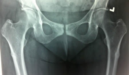 One prospective cohort study found that the risk of atypical fracture increased with longer duration of bisphosphonate use in women, particularly beyond 5 years of use.[46] This risk rapidly decreased after bisphosphonate discontinuation. The absolute risk of atypical femur fracture remained very low compared with reductions in the risk of hip and other fractures with bisphosphonate treatment although the risk-benefit balance appeared less favorable for Asian women.[46]
weak
chronic renal failure
Associated with a slightly increased fracture risk.[18]
diabetes mellitus
Associated with a slightly increased fracture risk.
male sex (acute fractures)
Males have a higher incidence of acute fractures.
female sex (fatigue and insufficiency fractures)
Females may have a higher risk for fatigue and insufficiency fractures.
Use of this content is subject to our disclaimer