Approach
Long bone fractures usually manifest with pain, swelling, and impaired function of the extremity. Deformity indicates a displaced fracture. The patient may guard any movement of the affected limb (e.g., during physical exam). Open fractures are often obvious, but sometimes an apparently minor surface wound belies severe injury below or nearby (bone can piston, leading to a wound near the site of injury). Therefore, any fracture associated with an overlying or nearby soft tissue injury, even an apparently innocuous minor wound, needs to be treated as an open fracture until shown otherwise.
Pathologic or insufficiency fractures may occur during seemingly low-energy stress.[1][53] The patient presents with sudden onset of pain, with rapid swelling, ecchymosis, and impaired function of the limb. Insufficiency fractures warrant a workup for metabolic bone disease. Discovery of a lytic lesion or other evidence of neoplasia requires an appropriate workup for malignancy.
Patients with dementia may exhibit withdrawal from attempted pressure or motion to the affected area, lack of use of the involved extremity, and nonspecific signs such as decreased appetite, new or worsened incontinence, or depression.
Signs indicating a vascular injury include lack of palpable pulse, continued blood loss, or expanding hematoma.[54] After x-rays, if a vascular injury is suspected, Doppler pressure (ankle/brachial systolic pressure index [ABI]), ultrasound duplex scanning, or CT angiography (if ABI is low) are often used to assess for vascular injury prior to angiography. In adults with multiple injuries, and complex or open fractures, a whole-body CT (including a vertex to toes scanogram followed by a vertex to mid-thigh CT scan) can be used.[54][55] The findings can direct further CT of the limbs as needed.[54]
The patient should be assessed for compartment syndrome (see Compartment syndrome of the extremities). In patients with long bone fractures, compartment syndrome is seen most commonly following fractures of the tibia or forearm.[56] The key clinical findings are pain out of proportion to the associated injury and pain on passive movement of the muscles of the involved compartments. The classic signs of acute compartment syndrome are loss of distal pulses, pallor, increased pain with passive stretch of tissues distal to fracture site, paresthesias, and poikilothermia. Negative predictive value is high, but the positive predictive value of these signs separately is low. A high index of suspicion is therefore needed since vascular injury without acute compartment syndrome can also cause pain, pallor, paresthesias, and loss of distal pulses.[57] Palpable pulses distal to the involved compartment do not rule out compartment syndrome. To recognize extremity compartment syndrome in a timely fashion, it is important to maintain a high index of suspicion and serially examine patients at risk to document changes over time. If the exam is equivocal or diagnosis is unclear, then there is a role for pressure measurement.[54][58] However, a "normal" compartment pressure alone should not be relied on to rule out acute compartment syndrome if the clinical picture is consistent with a compartment syndrome.
In patients with suspected stress fracture, radiographs are recommended initially but x-rays can be insensitive in the early stages of injury and in older patients with underlying osteoporosis.[1] If symptoms have been present for at least 10-14 days, radiographs are an effective screening tool since sensitivity reaches 30% to 70%.[1][59]
If radiographs confirm a stress fracture, magnetic resonance imaging (MRI) or CT of area of interest without intravenous contrast is usually appropriate to determine the extent of fracture or associated complications.[1]
In patients with suspected stress fractures and negative or indeterminate radiographs, MRI area of interest without intravenous contrast is usually appropriate, including in patients at high risk for fracture completion (e.g., patients with osteoporosis on bisphosphonate therapy).[1]
Bone scans of the whole body with single photon emission CT (SPECT) or SPECT/CT of the area of interest may also be indicated in patients with confirmed or suspected stress fractures.[1] Stress fractures are frequently evident days to weeks earlier on bone scans than radiographs. Bone scintigraphy can differentiate between osseous and soft tissue injury.[1] Normal bone scintigraphy generally excludes a diagnosis of stress fracture, and the patient can return to normal activity in most cases.[1] However, in older patients or patients with osteoporosis, abnormalities may not show up on bone scintigraphy for several days after the injury.[1] Patients using corticosteroids may also have less sensitive bone scintigraphy results.[1][60]
Humeral shaft fractures
These commonly result from direct trauma to the humerus and falls onto the outstretched hand.[24][25] Less commonly, extreme muscle contraction, electrocution injury, or seizure may lead to humeral shaft fracture.[26]
In displaced fractures, deformity is common, with the arm appearing shortened. Palpation reveals point tenderness at the fracture site, and bony crepitus is often palpable. A thorough neurovascular exam is required, as neurovascular compromise may occur. The radial nerve, and the brachial artery and vein are at greatest risk of compromise.
Plain radiographs of the humerus should include at least two 90° orthogonal views (anteroposterior [AP] and lateral), with the films encompassing the glenohumeral and elbow joints to evaluate for associated injury in these areas.
Fractures proximal to the pectoralis major insertion tend to result in the proximal fragment being internally rotated and abducted by rotator cuff forces. If the fracture is distal to the pectoralis major insertion but proximal to the deltoid insertion, the pull of the pectoralis major and arm adductors displace the proximal fragment medially, while the deltoid pulls the distal fragment laterally. Fractures distal to the deltoid insertion show abduction and flexion of the proximal fragment, with proximal displacement of the distal fragment.
Proximal humeral shaft fractures
These are typically seen in older people after a fall on the outstretched hand. Direct trauma and seizures may also lead to these fractures.[27] Most of these fractures are impacted; displacement and angulation are usually minimal. The surgical neck and greater tuberosity are the most common fracture sites.
Severe shoulder pain, swelling, and impaired range of motion may mimic the presentation of a shoulder dislocation. Deformity is uncommon unless associated dislocation is present, but swelling may make this hard to discern. Injury to the rest of the shoulder girdle may occur, so a focused yet thorough exam needs to be performed. Injury to the axillary nerve (tested by sensation over the regimental patch and deltoid and teres minor motor function), axillary artery, or circumflex humeral arteries may occur, so neurovascular exam is crucial.
X-rays should include views of the entire humerus, including the elbow, with anteroposterior (AP), axillary lateral, and scapular "Y" views to evaluate for fracture and dislocation. Any suspicion of more distal injury may require a separate elbow/forearm x-ray series. If vascular injury is suspected, CT angiogram (CTA) is indicated, along with orthopedic and vascular surgery consultation. CTA has largely supplanted the use of conventional angiography in assessing suspected vascular injury but angiography can be used in absence of CTA. CT or MRI may be needed for definitive diagnosis of subtle fractures (nondisplaced greater tuberosity fractures, sometimes even displaced lesser tuberosity fractures), for better evaluation of articular surface involvement in humeral head fractures, or for preoperative planning. Three-dimensional CT reconstruction has been advocated as a way to better visualize the exact anatomy of these injuries.[61]
Several classification systems exist for proximal humerus fractures, with the Neer system being the most widely used.[4] Newer classification systems have added articular surface orientation and other fracture characteristics.[5][6]
Humeral stress fractures
These are rare and primarily occur as a result of overuse among throwing athletes. Gymnasts, weightlifters, and other athletes who place repetitive high-impact or -torque loads on the humerus have also been known to sustain these injuries.
The person typically experiences gradually progressive arm pain with activity, but the completion of the fracture can be dramatic. Severe pain, swelling, and deformity of the shaft may be accompanied by neurovascular injury, especially to the radial nerve.[62][63] As such, it is important that an athlete complaining of persistent arm soreness be evaluated for a stress fracture before completion of the fracture occurs.
Plain films are often negative initially, so a triple-phase bone scan (TPBS) or MRI may be necessary to delineate the injury early on. Completed stress fractures may occur in throwing/overhead athletes (often with a prodrome of pain that the athlete has continued to push through).
Radial and ulnar shaft fractures
Radial shaft fractures usually result from a fall onto the outstretched or pronated wrist, or from a direct blow. Fractures at the junction of the mid and distal thirds of the shaft are often complicated by injury to the distal radioulnar joint; such injuries are often referred to as Galeazzi fractures. Because of the deforming muscle forces that act on these fractures, they are often dorsally angulated and are inherently unstable. The patient typically presents with pain and swelling over the mid to distal radius down into the wrist. Deformity may be noted. The fracture site and the distal radioulnar joint are tender to palpation. AP and lateral x-ray views should be ordered, ensuring that the entire radius and ulna, along with the wrist and elbow joints, are clearly visualized.
Isolated fractures of the mid shaft of the ulna, often called nightstick fractures, usually result from a person trying to ward off a blow from a heavy, blunt object (e.g., a night stick or truncheon). The patient typically presents with pain and swelling over the affected area. Point tenderness at the fracture site is noted, but deformity is uncommon. X-rays of the radius and ulna reveal the injury.
If the ulnar fracture involves the proximal third of the shaft, there may be associated dislocation of the radial head at the elbow (Monteggia fracture/dislocation). These fractures are rare and usually due to a fall onto the outstretched hand, with the elbow extended and pronated.[28] The patient presents with pain and swelling at the proximal ulna, with variable deformity depending on the exact nature of the injury. Associated injury to the radial nerve or posterior interosseous nerves may result in abnormal sensation at the dorsum of the thumb and second and third fingers. Wrist and thumb extension may also be weak. Plain films of the radius and ulna need to include the entire radius and ulna, along with the wrist and elbow joints. The radial head dislocation may be subtle, so it is important that the examiner closely inspects the radiographs for this injury. In the uninjured elbow, a line drawn through the axis of the proximal radial shaft and radial head (McLaughlin line) should bisect the capitellum.
Concomitant fractures of both the radius and the ulna are usually the result of high-energy trauma from a blow, fall, or motor vehicle accident. Pain and swelling at the fracture site are rarely accompanied by deformity. Injury to the radial, median, or ulnar nerves may occur, along with vascular injury.
The Monteggia and Orthopaedic Trauma Association (OTA) classification systems are commonly used for radial and ulnar fractures.[8][9][Figure caption and citation for the preceding image starts]: X-ray showing osteoporotic ulnar fracture involving the proximal third of the shaft with associated dislocation of the radial head at the elbow (Monteggia fracture)From the personal collection of Dr Philip H. Cohen [Citation ends].
Radial and ulnar stress fractures
These are uncommon and primarily occur among athletes who repetitively load the bones with high forces (e.g., gymnasts).[29][30] TPBS or MRI can show the fracture long before it becomes apparent on plain x-rays, and are usually recommended in higher-risk athletes with persistent and/or worsening pain, especially if the pain limits the athlete's activities.
Femoral shaft fractures
These are generally caused by high-energy trauma, such as a motor vehicle accident, or fall from a height. Spiral fractures of the femoral shaft may occur as a result of a twisting injury. Comminuted and open fractures may occur from gunshot wounds or other forms of high-energy penetrating trauma. Because great forces are required to break this large bone, femur fractures are often found in conjunction with other serious injuries. Therefore, the clinician must perform a diligent head-to-toe search for other signs of injury.
The patient with an acute femoral shaft fracture presents with severe pain of the involved extremity and is unable to walk. The thigh may appear markedly swollen, and deformity is often present. However, a large amount of blood can be lost into the thigh before it becomes clinically apparent, and patients can become hypotensive and go into hypovolemic shock from these injuries.[64] Thus, as part of the standard Advanced Trauma Life Support (ATLS)/Advanced Cardiac Life Support (ACLS) approach used for high-energy trauma patients, these patients should have their hemoglobin assessed with serial testing as indicated, and should be typed and cross-matched for transfusion. Frequent monitoring of vital signs is essential. Bruising of the thigh is often noted, and an expanding hematoma indicates severe vascular injury. Distal pulses may be weak or absent, as a result of either hypotension or acute compartment syndrome. Serial neurovascular exams are important to monitor for signs of developing compartment syndrome.
Once the patient has been hemodynamically stabilized (and appropriate analgesia has been given), plain AP and lateral radiographs of the femur need to be obtained. These fractures are usually significantly displaced and angulated.
There are several classification systems for femoral shaft fractures. One of the most common is the Winquist-Hansen system.[10][11][Figure caption and citation for the preceding image starts]: X-ray showing midshaft femur fractureFrom the personal collection of Dr Philip H. Cohen [Citation ends].
Femoral stress fractures
These generally have an indolent presentation. They typically manifest as pain with impact activity (e.g., running) that tends to get progressively worse over time. The pain is usually noticed in the groin or buttocks for a femoral neck stress fracture, along the thigh in a femoral shaft stress fracture, and around the knee in a distal femoral stress fracture. Some counterintuitive data have suggested that bisphosphonate use may actually increase the risk of femoral shaft stress fracture, while decreasing the risk of other fractures.[65][66]
The patient with a femoral stress fracture may have an antalgic gait, but gross inspection of the hip and thigh is usually unremarkable. Palpation rarely causes tenderness, although vague tenderness over the thigh may sometimes be mistaken for evidence of a muscle strain. A fulcrum test (levering the femoral shaft over the examiner's forearm, which is placed underneath the femoral shaft) may reproduce symptoms in a femoral neck or proximal shaft stress fracture. Similarly, passive internal rotation of the hip often reproduces pain in a patient with a femoral neck stress fracture. A hop test (hopping on one or both legs) usually produces pain as well. As these exam findings can be subtle, it is very important to maintain a high index of suspicion for these injuries.[67]
Initial evaluation should include AP and lateral x-rays of the femur. If the initial films are negative, repeat x-rays may subsequently demonstrate callus formation. TPBS and MRI are highly sensitive for detecting these injuries early on. MRI is much more specific than TPBS, does not expose the patient to ionizing radiation, and provides additional information about the surrounding tissues.[68] Patients suspected of having osteopenia/osteoporosis should undergo bone mineral density evaluation (i.e., dual-energy x-ray absorptiometry [DXA] scanning). However, if the DXA scan measures the femoral neck after bony healing has occurred, a falsely normal or even high bone mineral density may be reported. Thus, the contralateral femoral neck should be used for the DXA if possible.
Tibial shaft fractures
Tibial shaft fractures may result from direct trauma (usually causes transverse or comminuted fractures) or indirect twisting forces (usually causes spiral or oblique fractures).[31]
In an acute tibial shaft fracture, the patient presents with severe pain and is usually unable to bear weight. Massive swelling and ecchymosis may occur, and open fractures are relatively common.
A long leg splint should be applied for protection and comfort, and full-length AP and lateral x-rays of the tibia and fibula (including the knee and ankle) should be obtained.
Generally, surgical treatment of tibial fractures is considered if x-rays reveal greater than:[69]
5 mm of displacement in the AP plane or mediolaterally
10° of angulation in the AP plane
5° of angulation mediolaterally
10° of rotation.
Orthopedic consultation should be sought to consider the best choice for the individual patient.[70]
The AO Foundation system of tibial shaft fractures is the most widely used classification system.[12]
Tibial stress fractures
Stress fractures of the tibia are relatively common in impact athletes, such as runners and basketball players, and in military personnel.[32][33]
They generally present with gradually progressive pain with impact and weight-bearing activities. Eventually, they may also cause pain at rest. They most commonly occur at the posteromedial tibial border, with anterior tibial crest stress fractures occurring less often.
Inspection is usually unremarkable, but palpation reveals point tenderness at the fracture site. A healing stress fracture may be evinced by palpable callus. Pain upon tuning fork vibration in the area of the fracture is felt to be fairly specific but not very sensitive. Pain is reproduced by having the patient jog, hop, or, in more severe cases, walk. X-rays may be negative early in the course of the injury but later tend to show periosteal thickening, sclerosis, and callus formation.
TPBS and MRI are much more sensitive for early detection of these injuries, and MRI allows for more specific evaluation of marrow edema and the surrounding soft tissue.[71] With anterior tibial stress fractures, x-rays may reveal the "dreaded black line" - a horizontal radiolucency in the anterior tibial cortex. These injuries tend to heal more slowly than the more common posteromedial stress fractures, and they have a higher rate of nonunion.[72]
Simultaneous fracture of tibial and fibular shafts
High-energy trauma may result in simultaneous fracture of both the tibial and fibular shafts. These injuries are usually displaced and are often open.
The patient presents with severe pain, deformity, and is usually unable to bear weight. Massive swelling and skin wounds from fracture fragments may be noted. Acute compartment syndrome may occur, so repeated neurovascular exam is crucial.
A long leg splint should be applied and full tibia/fibula radiographs should be taken, including the knee and ankle joints. Emergency orthopedic consultation is required.[Figure caption and citation for the preceding image starts]: X-ray showing a segmental fracture of the tibia and fibulaFrom the personal collection of Dr Philip H. Cohen [Citation ends].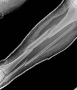
Isolated fibular fractures
These are typically caused by a direct blow to the outer aspect of the leg, or from an external rotation force at the ankle. In the latter situation, the force damages the distal tibiofibular syndesmosis, medial malleolus, and/or deltoid ligament of the ankle. The force is then transmitted proximally through the interosseous membrane and up the fibular shaft, until it blows out at the proximal fibula. This is referred to as a Maisonneuve fracture. In this case, the fibular fracture itself usually heals well with conservative management, but this should serve as a warning of a severe injury to the ankle structures (which generally require operative intervention).[73] See Ankle fractures.
The patient with a suspected fibular fracture needs to be evaluated for additional injuries, such as tibial fracture, ligamentous injury of the knee, and ankle fractures or instability. The common peroneal nerve runs over the proximal aspect of the fibula and may be injured by a proximal fibular fracture. This may lead to a foot drop and/or paresthesias at the lateral aspect of the leg and dorsum of the foot. With an isolated fibular shaft fracture, the patient typically has localized point tenderness, but dependent swelling and ecchymosis are often noted distally. The patient may be able to ambulate, albeit with discomfort.
AP and lateral x-rays of the full length of the tibia and fibula (including the knee and ankle joints) should be taken. More directed radiographic evaluation of the knee or ankle is indicated if the physical exam reveals evidence of injury at these locations.
Fibular stress fractures
Stress fractures of the fibula are uncommon but typically occur in runners and ballet dancers. Initial x-ray findings may be negative, but TPBS or MRI can demonstrate the fracture earlier.
Use of this content is subject to our disclaimer