Images and videos
Images


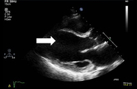

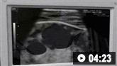
Acute heart failure
Diastolic image of dilated left ventricle (arrow)
From the private collections of Syed W. Yusuf, MBBS, MRCPI, and Daniel Lenihan, MD; used with permission
See this image in context in the following section/s:
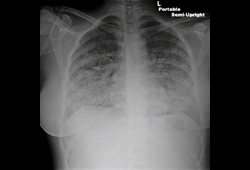
Acute heart failure
Chest x-ray of acute pulmonary edema showing increased alveolar markings, fluid in the horizontal fissure, and blunting of the costophrenic angles
From the private collections of Syed W. Yusuf, MBBS, MRCPI, and Daniel Lenihan, MD; used with permission
See this image in context in the following section/s:

Acute heart failure
Chest x-ray of acute pulmonary edema showing increased alveolar markings and bilateral pleural effusions
From the private collections of Syed W. Yusuf, MBBS, MRCPI, and Daniel Lenihan, MD; used with permission
See this image in context in the following section/s:
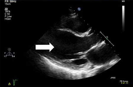
Acute heart failure
Systolic image of dilated left ventricle (arrow); note there is no change from diastolic image
From the private collections of Syed W. Yusuf, MBBS, MRCPI, and Daniel Lenihan, MD; used with permission
See this image in context in the following section/s:

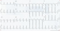
Acute heart failure
ECG showing left ventricular hypertrophy with sinus tachycardia
From the private collections of Syed W. Yusuf, MBBS, MRCPI, and Daniel Lenihan, MD; used with permission
See this image in context in the following section/s:
Videos
 Central venous catheter insertion: animated demonstration
Ultrasound-guided insertion of a non-tunnelled central venous catheter (CVC) into the right internal jugular vein using the Seldinger insertion technique.
 How to perform an ECG: animated demonstration
How to record an ECG. Demonstrates placement of chest and limb electrodes.
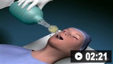 Tracheal intubation: animated demonstration
How to insert a tracheal tube in an adult using a laryngoscope.
Use of this content is subject to our disclaimer