Images and videos
Images
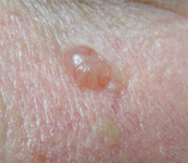


Basal cell carcinoma
Nodular basal cell carcinoma on the cheek, on background of diffuse solar damage with marked solar elastosis
From the collection of Prof. Robert A. Schwartz
See this image in context in the following section/s:
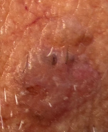
Basal cell carcinoma
Superficial BCC: A plaque somewhat translucent with focal crusting and ulceration, enlarging with a nodular quality
From the personal collection of Prof. Robert A. Schwartz; used with permission
See this image in context in the following section/s:

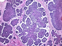
Basal cell carcinoma
Histologic appearance of basal cell carcinoma (20x, H-E stain); characteristic peripheral nuclear palisading, stroma-epithelium split, and so-called mucinous nature of the stroma are seen
From the collection of Drazen M. Jukic, MD, PhD
See this image in context in the following section/s:
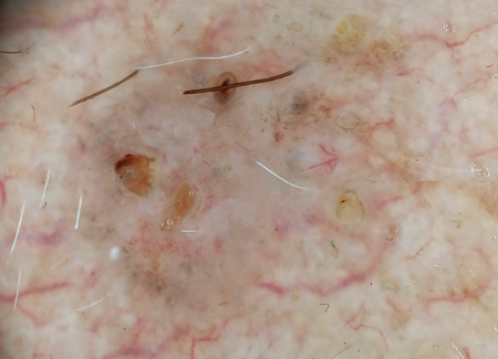
Basal cell carcinoma
Digital dermoscopy image of above superficial basal cell carcinoma utilizing Sklip PRO dermatoscope
From the personal collection of Prof. Robert A. Schwartz; used with permission
See this image in context in the following section/s:
Use of this content is subject to our disclaimer