Etiology
The etiology of basal cell carcinoma (BCC) is related to repetitive and frequent sun exposure, as ultraviolet (UV) radiation induces DNA damage in keratinocytes. There is exponential increase of BCCs in relation to UV exposure.[4][17] In particular, the 290-320 nanometer wavelength UV-B (sunburn wavelength) is believed to play an important role in the formation of BCC.[18] It should be viewed as a hazard to children and adolescents, as about one quarter of lifetime exposure occurs before 18 years of age.[19]
Pathophysiology
BCC development is the result of a complex interaction between environmental and genetic factors. BCCs arise from pluripotent cells in the epidermis and follicular epithelium.[20] In their attempt to recapitulate follicular and epidermal development, these cells progress to carcinomas. It is postulated that mutations of p53 gene may play a role in the development of BCC. In addition, aberrant activation of the Sonic Hedgehog signaling cascade contributes to the development of BCCs.[21]
Research to identify BCC cancer stem cells (tumor cells) is ongoing.[22][23]
Classification
Histopathology
The classification of BCC is a histopathologic one.[3][4][5][6] A biopsy to diagnose BCC is important to:
Determine histologic subtype (such as nodular, superficial, and pigmented), which may impact treatment, and
Exclude other skin cancers such as squamous cell carcinoma.
Nodular BCC
The most common clinical form of BCC, composed predominantly of a large nodule of mitotically-active hyperchromatic blue cells. Margins are easier to ascertain in this type. Macroscopically it is a pearly white nodule/tumor with prominent telangiectasia on its surface.[3][4][6][Figure caption and citation for the preceding image starts]: Nodular basal cell carcinoma on the cheek, on background of diffuse solar damage with marked solar elastosisFrom the collection of Prof. Robert A. Schwartz [Citation ends].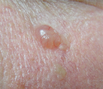
Superficial BCC
This variant clinically has poorly defined margins. On histopathologic examination, it is represented by small nests of atypical blue cells that seem to hang from the epidermis. The stroma surrounding these nests is more cellular than the rest of the dermis. The presence of this kind of stroma on the margin is considered a positive margin, meaning it is very likely there is more tumor in the patient's remaining skin tissue, and additional therapy is necessary.[6][7]
Micronodular BCC
Appears as multiple small nests in the dermis, overall well-circumscribed. It tends to be more likely to recur than nodular BCC, having intermediate characteristics between nodular and infiltrative subtypes.[8] Individual nests as well as the entire group are surrounded by cellular stroma, focally with myxoid features.[3][4]
Metatypical BCC (BCC with squamous differentiation)
Has a higher recurrence rate, and is therefore prognostically important. Any of the BCC types indicated above can also be metatypical (denoting BCC with squamous differentiation). Most notably, BCCs that have been repetitively rubbed undergo superficial squamous differentiation and should not be considered metatypical. Squamous differentiation needs to be present in the deep part of the neoplasm to diagnose such an entity.[3][4][9]
Morpheaform BCC
Synonymous with infiltrating or sclerosing BCC. Composed of infiltrating strands of mitotically active, hyperchromatic blue cells with prominent fibrotic (sclerotic) stroma. Clinically, this type is similar to plaques of morphea (scleroderma), hence the name. The margins of morpheaform BCC are less well-defined. Perineural invasion is common; therefore, local recurrences are more often seen than with other types.[3][4][6]
Infundibulocystic BCC
So named as it commonly simulates trichoepithelioma, a benign tumor with follicular differentiation. Infundibulocystic BCC is composed of strands and nests of mitotically active hyperchromatic blue cells, with formation of so-called keratinous cysts, which represent an attempt of BCC to form superficial keratin. The chief significance is in the frequent misdiagnosis of this type as trichoepithelioma or trichoblastoma group of neoplasms.[3][4]
Mixed BCC
Most often (with exception of superficial BCC) a pure subtype of BCC will not be observed. Usually there is a mixture of two or more of the types described above. It is important to denote the subtype of BCC in the pathologic report, as clinical treatment will depend on it. It is worth noting that the longer a BCC is present, the more likely bleeding, ulceration, or crusting will be present.[3][4]
Periocular BCC
Usually a slow developing, painless and non-resolving lesion. The typical form and location is a small hard whitish nodule that appears on the lower eyelid. Periocular BCC can also present in the medial canthus, upper lid, and lateral canthus.[10]
It can present as nodular (hard nodule, pearly appearance, abnormal [telangiectatic] vessels), nodulo-ulcerative (as nodular but with raised rolled border surrounding central ulcer, may bleed), or morphoeic or sclerosing (flat hardened plaque of thickened skin, without surface vascularization, ill-defined border making it difficult to determine area of involvement).
Use of this content is subject to our disclaimer