Differentials
Condyloma latum
SIGNS / SYMPTOMS
One of the manifestations of secondary syphilis.
Patients may have associated systemic signs and symptoms, such as fever, malaise, adenopathy, and weight loss.
The lesions appear as moist, whitish, wart-like papules, and are highly contagious.[Figure caption and citation for the preceding image starts]: Condylomata lata: this photograph depicts the appearance of what was determined to be secondary syphilitic lesions, also known as "condylomata lata," on a patient’s perineumCDC/Joyce Ayers [Citation ends].
Lesions relapse in approximately 20% of patients without treatment within 1 year.[3][45]
INVESTIGATIONS
Direct detection of treponemes in specimen under dark-field microscopy is diagnostic.
Serologic tests for syphilis, such as rapid plasma reagin, may be helpful.
Molluscum contagiosum
SIGNS / SYMPTOMS
A viral infection of the epidermis that manifests as flesh-colored papules that are often umbilicated. [Figure caption and citation for the preceding image starts]: Extensive molluscum lesions on the flank of a young child; lesions are flesh- to pearly-colored with central dellsFrom the personal collection of Nanette Silverberg, Beth Israel Medical Center, NY; used with permission [Citation ends].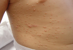
The infection is self-limited, and lesions persist up to 6 months.
Occurs in children and sexually active adults. In children, the lesions are seen on exposed skin, but in sexually active adults, lesions occur in the genital region.
In HIV-positive patients, the clinical presentation is generally more severe with a greater number of lesions and larger lesions that may involve the face.
These lesions are usually asymptomatic unless secondarily infected, in which case they may be painful.[3][46]
INVESTIGATIONS
Diagnosis is usually clinical. However, direct microscopic examination of Giemsa-stained central core shows molluscum or inclusion bodies.
Biopsy may be indicated in HIV-infected patients to rule out fungal infection.
Pearly penile papules
SIGNS / SYMPTOMS
Pearly penile papules are a normal anatomic variant of the glans that appear as flesh-colored, discrete papules approximately 1 to 2 mm in size, distributed evenly around the corona.
They are asymptomatic but may generate anxiety in the patient.[3][46]
INVESTIGATIONS
Diagnosis is clinical.
Hidradenoma papilliferum
Seborrheic keratosis
SIGNS / SYMPTOMS
Common, benign verrucous lesions that may vary in color from tan to brown to gray to black. They have a classic "stuck-on" appearance.[Figure caption and citation for the preceding image starts]: Clinical close-up image of seborrheic keratosis on the back of a 40-year-old manFrom the collection of Dr Ralph Braun, University Hospital Zurich and Dr Isabel Kolm, University Hospital Zurich; used with permission [Citation ends].
They evolve over months to years and rarely occur before the age of 30.[3][46]
The lesions tend to appear on the face, torso, and upper extremities.
INVESTIGATIONS
Diagnosis is clinical.
Carcinoma in situ
SIGNS / SYMPTOMS
May appear as multifocal erythematous macules, lichenoid, or pigmented papules sometimes forming plaques on the external anogenital region. The surface is usually smooth and velvety.
Ulceration suggests invasiveness. Cervix and anus involvement have the highest risk to become invasive.
INVESTIGATIONS
Diagnosis is clinical; however, it should always be confirmed by biopsy.
Dermatopathology shows epidermal proliferation with numerous abnormal mitoses, dyskeratotic cells, and atypical pleomorphic cells with large hyperchromatic nuclei.[46]
Bowenoid papulosis/erythroplasia of Queyrat
SIGNS / SYMPTOMS
Bowenoid papulosis can appear as red-brown papules that may coalesce to form a plaque. These can often closely resemble benign condylomata.
Erythroplasia of Queyrat typically appears as as erythematous, well-defined, velvety plaque of the glans or vulva.[5]
INVESTIGATIONS
Atypical proliferating suprabasal cells present in the full thickness of the epithelium.[46]
Skin tags
SIGNS / SYMPTOMS
Soft, skin-colored or tan-brown, round-oval, pedunculated papilloma.
Normally constricted at the base, and the size varies from 1 mm to 1 cm.
INVESTIGATIONS
Diagnosis is clinical.[46]
Use of this content is subject to our disclaimer