Tests
1st tests to order
blood cultures
Test
At least three sets of blood cultures from different venipuncture sites should be taken, the first and last samples taken at least one hour apart (ideally >6 hours apart if clinical status allows).[6][7] Blood culture results taken this way are positive in 90% of patients with IE.[20] Cultures should not be taken from indwelling lines, to minimize risk of contamination.
However, empiric antibiotic therapy should not be delayed while waiting to take three sets of blood cultures if the patient is unwell (e.g., with sepsis).[64] The volume of blood sent for culture is more important than the number of sets of cultures.[65] The most common cause of culture-negative endocarditis is antibiotic therapy preceding blood cultures.[66] Do not routinely request extended incubation of blood cultures in suspected endocarditis.[20][67]
Result
bacteremia; fungemia
echocardiogram
Test
Should be performed in all cases of suspected IE as early as possible to confirm or rule out the diagnosis.[6][7][62][63][75] Echocardiography is also important for evaluation of complications and prognosis.[62][Figure caption and citation for the preceding image starts]: A transthoracic echo showing large mobile vegetations on the anterior and posterior leaflets of the tricuspid valveFoley JA, Augustine D, Bond R, et al. Lost without Occam's razor: Escherichia coli tricuspid valve endocarditis in a non-intravenous drug user. BMJ Case Rep. 2010 Aug 10;2010. pii: bcr0220102769 [Citation ends]. [Figure caption and citation for the preceding image starts]: Image from transesophageal echocardiogram. White arrow indicates vegetation on the patient's aortic valveTeoh LS, Hart HH, Soh MC, et al. Bartonella henselae aortic valve endocarditis mimicking systemic vasculitis. BMJ Case Rep. 2010 Oct 21;2010. pii: bcr0420102945 [Citation ends].
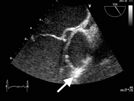
The decision to obtain a transthoracic echocardiogram (TTE) rather than transesophageal echocardiogram (TEE) is often difficult and has been considered by various guidelines.[6][7][62][63][75] The American Heart Association and the American College of Cardiology recommend that any patient suspected of having native valve IE should be screened with TTE to evaluate for the presence of a vegetation, and to assess any effects on valvular function.[20]
TEE is the preferred form of echocardiography in people with prosthetic material suspected to have IE.[63] TEE is also indicated in patients with a positive TTE, but where complications are suspected or likely, and before cardiac surgery during active IE.
Result
valvular, mobile vegetations
CBC
Test
Most patients have a normocytic, normochromic anemia. Leukocytosis is seen in about one third of cases, often with neutrophilia.
Result
anemia; leukocytosis
CRP
Test
Nonspecific test. High in almost all patients.[86]
In practice, CRP is useful as a baseline test and to monitor response to treatment.
Result
markedly elevated
serum chemistry panel with glucose
Test
Provides baseline assessment.
Result
normal or elevated BUN
LFTs
Test
Provides baseline assessment.
Result
normal or elevated
urinalysis
Test
Septic emboli are common complications of IE, and urinalysis may demonstrate active sediment assisting in the clinical diagnosis.
Result
RBC casts; WBC casts; proteinuria; pyuria
ECG
Test
Progression of the infection may lead to conduction system disease.[72]
Conduction abnormalities secondary to IE (mainly first-, second-, and third-degree atrioventricular block) are uncommon but are associated with worse prognosis and higher mortality compared with patients without conduction abnormalities.[73]
Result
prolonged PR interval; nonspecific ST/T wave abnormalities; atrioventricular block
Tests to consider
rheumatoid factor
erythrocyte sedimentation rate
Test
Nonspecific test. High in almost all patients.
Result
markedly elevated
complement levels
Test
May be performed.
Result
decreased
cardiac CT
Test
CT imaging is used for the diagnosis of native and prosthetic valve IE and for detection of complications of IE, including abscesses, pseudoaneurysms, and fistulae.[74][75] Cardiac CT has been found to compare favorably with transthoracic echocardiogram in detecting valvular abnormalities in patients with IE, but may miss small defects (e.g., small leaflet perforations [≤2 mm diameter], small vegetations [<10 mm]).[7][76]
Whole-body and brain CT can be used to look for distant lesions, systemic complications of IE, and septic emboli, as well as having a role in detecting extracardiac sources of bacteremia and potentially alternate diagnoses in patients where IE has been excluded.[6][7] CT angiography is a sensitive and specific test for detecting mycotic arterial aneurysms; MRI is superior for assessment of neurologic complications in terms of imaging, but is limited by accessibility and availability.[6][7]
Result
valvular abnormalities and vegetations
MRI
Test
MRI is of less diagnostic value that CT, but is the imaging modality of choice when investigating the cerebral complications of IE, with studies consistently reporting cerebral infarcts in up to 80% of patients.[77][78] MRI also reveals cerebral lesions in 50% of patients who do not demonstrate neurologic symptoms.[79] MRI is also used to assess spinal involvement in IE, including spondylodiscitis and vertebral osteomyelitis.[7]
Result
may show cerebral lesions, spinal involvement
Nuclear imaging and PET
Test
Single-PET/CT and 18F-fluorodeoxyglucose PET/CT may be particularly useful in patients with “possible IE” defined according to Duke criteria, in the demonstration of infectious embolic events, and in cases of suspected prosthetic valve IE where echocardiography is not diagnostic.[20][75][80][81][84]
Whole-body imaging with 18F-FDG-PET/CT can be useful to detect distant lesions, mycotic aneurysms, and portal of entry of bacteria, and to monitor response to antibiotic treatment in patients for whom surgery is being considered.[7][82][84]
Result
high level of local inflammation independently associated with a high risk of new‐onset embolic events; may reveal distant lesions, mycotic aneurysms, portal of bacterial entry; used to monitor response to antibiotic treatment
Emerging tests
mean platelet volume (MPV)
Test
MPV is associated with platelet activation which occurs in the setting of endothelial damage. Increased MPV has been shown to be an independent predictor of embolic events in patients with IE.[68]
Result
may be elevated; >8.6 femtoliters strongly predictive of embolic events
anti‐beta-2‐glycoprotein I antibodies
Test
Anti‐beta-2‐glycoprotein I antibodies enhance activation of platelets and the coagulation cascade and have been associated with increased risk of embolic events.[68]
Result
may be elevated
D-dimer and troponin I
Test
D-dimer and troponin I have also been linked to increased embolic risk but are nonspecific and may be surrogate markers for severity of illness rather than independent predictors of embolic events.[68]
Result
may be elevated
Use of this content is subject to our disclaimer