Images and videos
Images


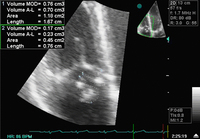


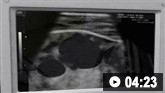
Infective endocarditis
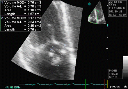
A transthoracic echo showing large mobile vegetations on the anterior and posterior leaflets of the tricuspid valve
Foley JA, Augustine D, Bond R, et al. Lost without Occam's razor: Escherichia coli tricuspid valve endocarditis in a non-intravenous drug user. BMJ Case Rep. 2010 Aug 10;2010. pii: bcr0220102769
See this image in context in the following section/s:

Infective endocarditis
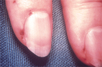
Osler node
From the collection of Sanjay Sharma, St George’s University of London, UK; used with permission
See this image in context in the following section/s:
Infective endocarditis
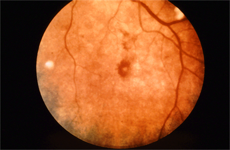
Roth spots
From the collection of Sanjay Sharma, St George’s University of London, UK; used with permission
See this image in context in the following section/s:

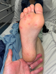
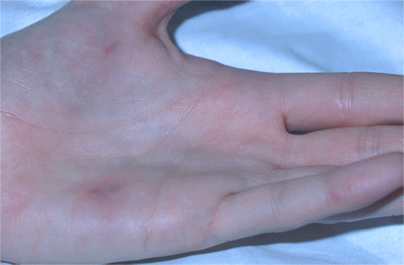
Infective endocarditis
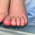
Cutaneous infarcts
From the collection of Sanjay Sharma, St George’s University of London, UK; used with permission
See this image in context in the following section/s:

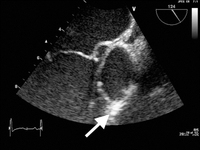
Infective endocarditis
Image from transesophageal echocardiogram. White arrow indicates vegetation on the patient's aortic valve
Teoh LS, Hart HH, Soh MC, et al. Bartonella henselae aortic valve endocarditis mimicking systemic vasculitis. BMJ Case Rep. 2010 Oct 21;2010. pii: bcr0420102945
See this image in context in the following section/s:

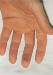
Infective endocarditis
Osler's nodes
From a private collection; used with permission
See this image in context in the following section/s:
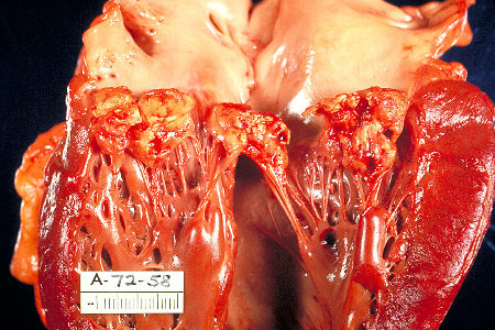
Infective endocarditis
Gross pathology of subacute bacterial endocarditis involving mitral valve
CDC/Dr Edwin P. Ewing, Jr.; used with permission
See this image in context in the following section/s:
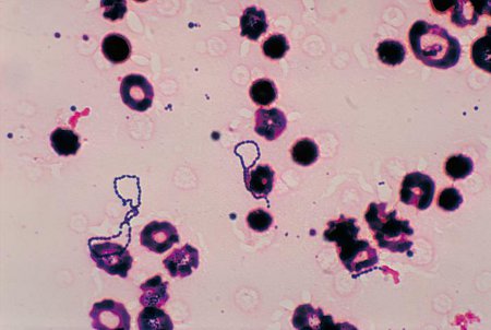
Infective endocarditis
Photomicrograph of Streptococcus viridans bacteria that had been grown in a blood culture
CDC/Dr Mike Miller; used with permission
See this image in context in the following section/s:
Infective endocarditis
Janeway lesions
From the collection of Sanjay Sharma, St George’s University of London, UK; used with permission
See this image in context in the following section/s:

Infective endocarditis
Janeway lesions
From a private collection; used with permission
See this image in context in the following section/s:
Videos
 Venepuncture and phlebotomy: animated demonstration
How to take a venous blood sample from the antecubital fossa using a vacuum needle.
 How to perform an ECG: animated demonstration
How to record an ECG. Demonstrates placement of chest and limb electrodes.
 Peripheral intravascular catheter: animated demonstration
How to insert a peripheral intravascular catheter into the dorsum of the hand.
 Central venous catheter insertion: animated demonstration
Ultrasound-guided insertion of a non-tunnelled central venous catheter (CVC) into the right internal jugular vein using the Seldinger insertion technique.
Use of this content is subject to our disclaimer