Images and videos
Images


Hereditary spherocytosis
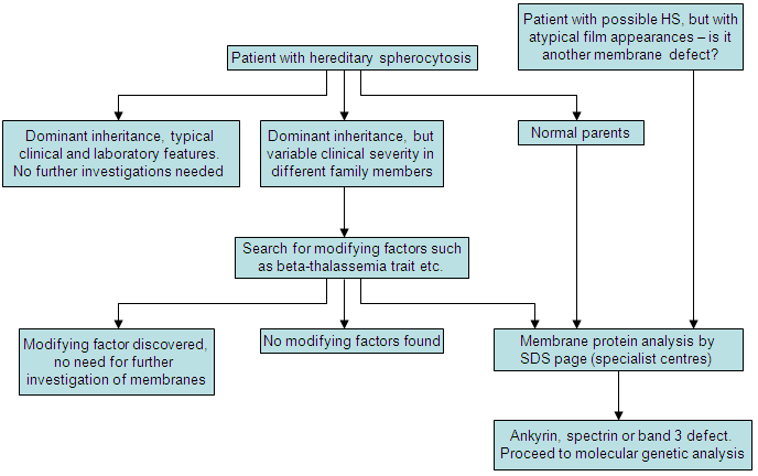
Diagnostic algorithm for HS
Created by the BMJ Evidence Centre using information from author
See this image in context in the following section/s:

Hereditary spherocytosis
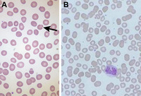
Blood smear of patient with HS (A) compared with normal blood smear (B); Pincer cell (mushroom-shaped cell) indicated
From the collection of Paula Bolton-Maggs, University of Manchester, UK; used with permission
See this image in context in the following section/s:
Hereditary spherocytosis
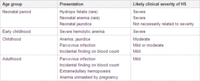
Presentation of HS by age
Created by the BMJ Evidence Centre using information from author
See this image in context in the following section/s:
Hereditary spherocytosis
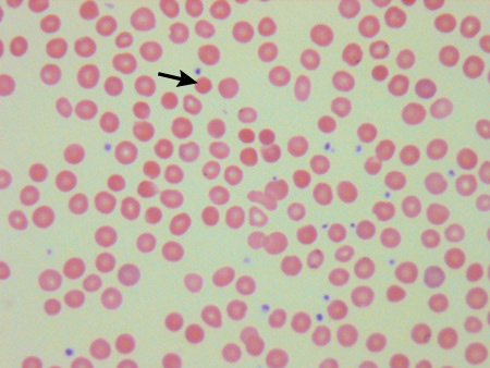
Blood smear of a patient with HS; spherocyte indicated
From the collection of Shelley Crary, University of Texas Southwestern Medical Center, TX; used with permission
See this image in context in the following section/s:
Hereditary spherocytosis
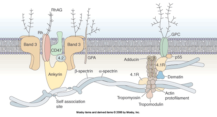
A schematic model of the organization of the red-cell membrane (the proteins and lipids are not drawn to scale)
This figure was published in: Young NS, Gerson SL, High KA, eds. Clinical hematology. Mohandas N, Reid ME, Erythrocyte structure, pp 36-38. Copyright Mosby Elsevier; 2006
See this image in context in the following section/s:
Hereditary spherocytosis
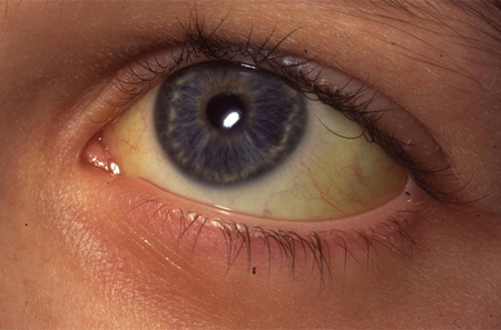
Jaundiced sclera in eye of child with HS
From the collection of Paula Bolton-Maggs, University of Manchester, UK; used with permission
See this image in context in the following section/s:
Use of this content is subject to our disclaimer