Images and videos
Images


Mastitis and breast abscess
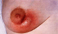
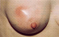
A breast abscess that developed during breast-feeding
From the collection of Mr R. Vashisht, West Middlesex University Hospital, London; used with permission
See this image in context in the following section/s:

Mastitis and breast abscess
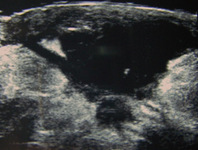
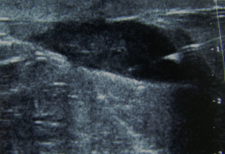
Ultrasound image showing a well-circumscribed hypoechoic breast abscess
From the collection of Holly S. Mason, MD, Tufts University School of Medicine, MA
See this image in context in the following section/s:

Mastitis and breast abscess
Nonlactating breast abscess due to periductal mastitis
From the collection of Mr R. Vashisht, West Middlesex University Hospital, London; used with permission
See this image in context in the following section/s:
Mastitis and breast abscess
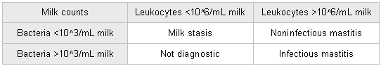
Leukocyte counts and bacteria quantification in breast milk
Chart produced by author using data from Thomsen AC, Espersen T, Maigaard S. Am J Obstet Gynecol. 1984 Jul 1;149(5):492-5
See this image in context in the following section/s:
Mastitis and breast abscess
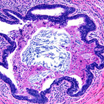
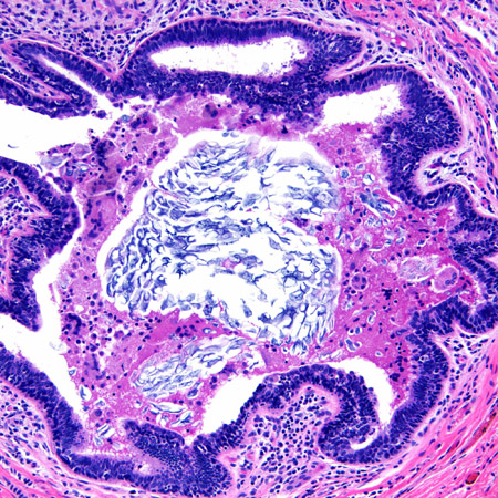
Duct ectasia with a central calcified keratin plug and associated giant cell inflammatory response
From the collection of Liron Pantanowitz, MD, Tufts University School of Medicine, MA
See this image in context in the following section/s:
Mastitis and breast abscess
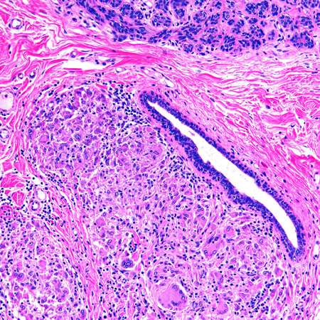
Microscopy image of non-necrotizing granulomatous inflammation in the breast
From the collection of Liron Pantanowitz, MD, Tufts University School of Medicine, MA
See this image in context in the following section/s:
Mastitis and breast abscess
Needle aspiration of a breast abscess under ultrasound guidance: note the needle piercing the abscess to the right of the image
From the collection of Holly S. Mason, MD, Tufts University School of Medicine, MA
See this image in context in the following section/s:
Mastitis and breast abscess
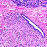
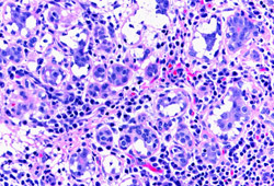
Lactational mastitis: microscopy image showing hypersecretory glands associated with inflammation
From the collection of Liron Pantanowitz, MD, Tufts University School of Medicine, MA
See this image in context in the following section/s:

Mastitis and breast abscess
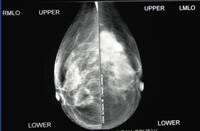
Tubercular mastitis: mammogram showing a mass lesion in the upper outer quadrant
Adapted from the Internet J Surgery (2007); used with permission
See this image in context in the following section/s:

Mastitis and breast abscess
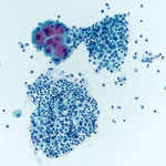
Aspirated material: a group of benign apocrine cells associated with acute and chronic inflammation in keeping with a breast abscess (ThinPrep stain)
From the collection of Liron Pantanowitz, MD, Tufts University School of Medicine, MA
See this image in context in the following section/s:
Use of this content is subject to our disclaimer