Intracerebral hemorrhage is a medical emergency that requires immediate attention.[70]Cordonnier C, Demchuk A, Ziai W, et al. Intracerebral haemorrhage: current approaches to acute management. Lancet. 2018 Oct 6;392(10154):1257-68.
http://www.ncbi.nlm.nih.gov/pubmed/30319113?tool=bestpractice.com
Arresting hematoma growth is a key objective for medical or surgical therapies.[21]Al-Shahi Salman R, Frantzias J, Lee RJ, et al. Absolute risk and predictors of the growth of acute spontaneous intracerebral haemorrhage: a systematic review and meta-analysis of individual patient data. Lancet Neurol. 2018 Oct;17(10):885-94.
https://www.thelancet.com/journals/laneur/article/PIIS1474-4422(18)30253-9/fulltext
http://www.ncbi.nlm.nih.gov/pubmed/30120039?tool=bestpractice.com
Initial stabilization and placement
Initial evaluation and management focuses on stabilization of airway, breathing, and circulation.[9]Greenberg SM, Ziai WC, Cordonnier C, et al. 2022 Guideline for the management of patients with spontaneous intracerebral hemorrhage: a guideline from the American Heart Association/American Stroke Association. Stroke. 2022 Jul;53(7):e282-361.
https://www.ahajournals.org/doi/full/10.1161/STR.0000000000000407
http://www.ncbi.nlm.nih.gov/pubmed/35579034?tool=bestpractice.com
For patients who are unable to protect their airway or who present with depressed level of consciousness (Glasgow Coma Score [GCS] ≤8), endotracheal intubation for airway protection is recommended.
Blood pressure (BP) is frequently elevated at presentation and requires treatment. Maintaining a systolic BP goal range of 130 to 150 mmHg is reasonable. Careful titration is recommended, to ensure continuous smooth and sustained control of BP, avoiding peaks and large variability in systolic BP.[9]Greenberg SM, Ziai WC, Cordonnier C, et al. 2022 Guideline for the management of patients with spontaneous intracerebral hemorrhage: a guideline from the American Heart Association/American Stroke Association. Stroke. 2022 Jul;53(7):e282-361.
https://www.ahajournals.org/doi/full/10.1161/STR.0000000000000407
http://www.ncbi.nlm.nih.gov/pubmed/35579034?tool=bestpractice.com
All patients with acute hemorrhagic stroke should be admitted to a neuroscience intensive care unit (ICU) because of the following potential risks or requirements:
Hourly neurologic vital signs
Intubation with mechanical ventilation
Depressed consciousness
High risk for hematoma expansion
BP monitoring or control with continuous infusions
Need for external ventriculostomy catheter, intracranial pressure (ICP) monitor, or surgical intervention.
Some patients require urgent placement of an external ventriculostomy catheter due to acute hydrocephalus.
Regardless of bleed location (cerebellar versus noncerebellar), neurologically stable patients with strong vital signs may be observed, ensuring adequate hydration and nutrition as necessary.
Evaluation and treatment are best accomplished in the neuroscience ICU, or eventually in a dedicated stroke unit.[9]Greenberg SM, Ziai WC, Cordonnier C, et al. 2022 Guideline for the management of patients with spontaneous intracerebral hemorrhage: a guideline from the American Heart Association/American Stroke Association. Stroke. 2022 Jul;53(7):e282-361.
https://www.ahajournals.org/doi/full/10.1161/STR.0000000000000407
http://www.ncbi.nlm.nih.gov/pubmed/35579034?tool=bestpractice.com
[101]Adeoye O, Nyström KV, Yavagal DR, et al. Recommendations for the establishment of stroke systems of care: a 2019 update. Stroke. 2019 Jul;50(7):e187-210.
https://www.ahajournals.org/doi/10.1161/STR.0000000000000173
http://www.ncbi.nlm.nih.gov/pubmed/31104615?tool=bestpractice.com
[102]Langhorne P, Ramachandra S, Stroke Unit Trialists' Collaboration. Organised inpatient (stroke unit) care for stroke: network meta-analysis. Cochrane Database Syst Rev. 2020 Apr 23;(4):CD000197.
https://www.cochranelibrary.com/cdsr/doi/10.1002/14651858.CD000197.pub4/full
http://www.ncbi.nlm.nih.gov/pubmed/32324916?tool=bestpractice.com
[  ]
How does organized inpatient care compare with care on a general medical ward for people with stroke?/cca.html?targetUrl=https://www.cochranelibrary.com/cca/doi/10.1002/cca.3108/fullShow me the answer
]
How does organized inpatient care compare with care on a general medical ward for people with stroke?/cca.html?targetUrl=https://www.cochranelibrary.com/cca/doi/10.1002/cca.3108/fullShow me the answer
Monitoring of all patients in a facility with 24-hour access to emergency neurosurgery coverage is recommended, in case of acute deterioration from hydrocephalus or mass effect. If 24-hour neurosurgery coverage is not available, the patient should be transferred to a hospital that has such coverage.
Surgical intervention
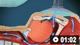
Surgical hematoma evacuation through craniotomy, minimally invasive approaches, or ventriculostomy is aimed at preventing further pressure-related injury and protecting against secondary physiological and cellular injury.[9]Greenberg SM, Ziai WC, Cordonnier C, et al. 2022 Guideline for the management of patients with spontaneous intracerebral hemorrhage: a guideline from the American Heart Association/American Stroke Association. Stroke. 2022 Jul;53(7):e282-361.
https://www.ahajournals.org/doi/full/10.1161/STR.0000000000000407
http://www.ncbi.nlm.nih.gov/pubmed/35579034?tool=bestpractice.com
Cerebellar hemorrhage
For patients with cerebellar intracerebral hemorrhage who are deteriorating neurologically, or have brainstem compression, or hydrocephalus from ventricular obstruction, or cerebellar intracerebral hemorrhage volume ≥15 mL, the American Heart Association and American Stroke Association (AHA/ASA) recommend immediate surgical removal of the hemorrhage with or without external ventricular drainage (EVD). Surgical intervention in these patients has been shown to reduce mortality, compared with medical management alone. The efficacy of surgical evacuation for improving functional outcomes, however, is uncertain and has not been demonstrated in retrospective studies.[9]Greenberg SM, Ziai WC, Cordonnier C, et al. 2022 Guideline for the management of patients with spontaneous intracerebral hemorrhage: a guideline from the American Heart Association/American Stroke Association. Stroke. 2022 Jul;53(7):e282-361.
https://www.ahajournals.org/doi/full/10.1161/STR.0000000000000407
http://www.ncbi.nlm.nih.gov/pubmed/35579034?tool=bestpractice.com
Noncerebellar hemorrhage
Limited data suggest that in patients with supratentorial intracerebral hemorrhage who are deteriorating, craniotomy for hematoma evacuation might be considered as a lifesaving measure.[9]Greenberg SM, Ziai WC, Cordonnier C, et al. 2022 Guideline for the management of patients with spontaneous intracerebral hemorrhage: a guideline from the American Heart Association/American Stroke Association. Stroke. 2022 Jul;53(7):e282-361.
https://www.ahajournals.org/doi/full/10.1161/STR.0000000000000407
http://www.ncbi.nlm.nih.gov/pubmed/35579034?tool=bestpractice.com
However, randomized trials comparing standard surgery (craniotomy) with conservative management have not demonstrated a clear benefit for surgical intervention in patients with intracerebral hemorrhage.[9]Greenberg SM, Ziai WC, Cordonnier C, et al. 2022 Guideline for the management of patients with spontaneous intracerebral hemorrhage: a guideline from the American Heart Association/American Stroke Association. Stroke. 2022 Jul;53(7):e282-361.
https://www.ahajournals.org/doi/full/10.1161/STR.0000000000000407
http://www.ncbi.nlm.nih.gov/pubmed/35579034?tool=bestpractice.com
[106]Mendelow AD, Gregson BA, Fernandes HM, et al. Early surgery versus initial conservative treatment in patients with spontaneous supratentorial intracerebral haematomas in the International Surgical Trial in Intracerebral Haemorrhage (STICH): a randomised trial. Lancet. 2005 Jan 29-Feb 4;365(9457):387-97.
http://www.ncbi.nlm.nih.gov/pubmed/15680453?tool=bestpractice.com
[107]Mendelow AD, Gregson BA, Rowan EN, et al; STICH II Investigators. Early surgery versus initial conservative treatment in patients with spontaneous supratentorial lobar intracerebral haematomas (STICH II): a randomised trial. Lancet. 2013 Aug 3;382(9890):397-408.
http://www.ncbi.nlm.nih.gov/pubmed/23726393?tool=bestpractice.com
Minimally invasive surgery for intracerebral hemorrhage
Recommendations from the AHA/ASA include the following:[9]Greenberg SM, Ziai WC, Cordonnier C, et al. 2022 Guideline for the management of patients with spontaneous intracerebral hemorrhage: a guideline from the American Heart Association/American Stroke Association. Stroke. 2022 Jul;53(7):e282-361.
https://www.ahajournals.org/doi/full/10.1161/STR.0000000000000407
http://www.ncbi.nlm.nih.gov/pubmed/35579034?tool=bestpractice.com
For patients with supratentorial intracerebral hemorrhage of 20- to 30-mL volume with GCS scores in the moderate range (5-12), minimally invasive hematoma evacuation with endoscopic or stereotactic aspiration with or without thrombolytic use can be useful to reduce mortality compared with medical management alone.
For patients with supratentorial intracerebral hemorrhage of 20- to 30-mL volume with GCS scores in the moderate range (5-12) being considered for hematoma evacuation, it may be reasonable to select minimally invasive hematoma evacuation over conventional craniotomy to improve functional outcomes.
For patients with supratentorial intracerebral hemorrhage of 20- to 30-mL volume with GCS scores in the moderate range (5-12), the effectiveness of minimally invasive hematoma evacuation with endoscopic or stereotactic aspiration with or without thrombolytic use to improve functional outcomes is uncertain.
Minimally invasive surgical interventions require surgeon and center skill and experience as the basis for these recommendations. The literature supports that minimally invasive surgery may be considered to improve functional outcomes compared with conventional craniotomy.[9]Greenberg SM, Ziai WC, Cordonnier C, et al. 2022 Guideline for the management of patients with spontaneous intracerebral hemorrhage: a guideline from the American Heart Association/American Stroke Association. Stroke. 2022 Jul;53(7):e282-361.
https://www.ahajournals.org/doi/full/10.1161/STR.0000000000000407
http://www.ncbi.nlm.nih.gov/pubmed/35579034?tool=bestpractice.com
However, the mortality benefit of minimally invasive surgery compared with craniotomy is uncertain since results from large randomized clinical trials have not been definitive.[9]Greenberg SM, Ziai WC, Cordonnier C, et al. 2022 Guideline for the management of patients with spontaneous intracerebral hemorrhage: a guideline from the American Heart Association/American Stroke Association. Stroke. 2022 Jul;53(7):e282-361.
https://www.ahajournals.org/doi/full/10.1161/STR.0000000000000407
http://www.ncbi.nlm.nih.gov/pubmed/35579034?tool=bestpractice.com
Systematic reviews and meta-analyses have shown that patients subject to early evacuation using minimally invasive techniques (within 24 hours) are more likely to achieve functional independence.[108]Scaggiante J, Zhang X, Mocco J, et al. Minimally invasive surgery for intracerebral hemorrhage: an updated meta-analysis of randomized controlled trials. Stroke. 2018 Nov;49(11):2612-20.
https://www.ahajournals.org/doi/10.1161/STROKEAHA.118.020688
http://www.ncbi.nlm.nih.gov/pubmed/30355183?tool=bestpractice.com
[109]Tang Y, Yin F, Fu D, et al. Efficacy and safety of minimal invasive surgery treatment in hypertensive intracerebral hemorrhage: a systematic review and meta-analysis. BMC Neurol. 2018 Sep 3;18(1):136.
https://bmcneurol.biomedcentral.com/articles/10.1186/s12883-018-1138-9
http://www.ncbi.nlm.nih.gov/pubmed/30176811?tool=bestpractice.com
[110]Xia Z, Wu X, Li J, et al. Minimally invasive surgery is superior to conventional craniotomy in patients with spontaneous supratentorial intracerebral hemorrhage: a systematic review and meta-analysis. World Neurosurg. 2018 Jul;115:266-73.
http://www.ncbi.nlm.nih.gov/pubmed/29730105?tool=bestpractice.com
[111]Cavallo C, Zhao X, Abou-Al-Shaar H, et al. Minimally invasive approaches for the evacuation of intracerebral hemorrhage: a systematic review. J Neurosurg Sci. 2018 Dec;62(6):718-33.
http://www.ncbi.nlm.nih.gov/pubmed/30160081?tool=bestpractice.com
One meta-analysis suggested that patients with superficial hematoma between 25 mL and 40 mL, treated within 72 hours, are most likely to benefit from a minimally invasive surgical approach.[112]Zhou X, Chen J, Li Q, et al. Minimally invasive surgery for spontaneous supratentorial intracerebral hemorrhage: a meta-analysis of randomized controlled trials. Stroke. 2012 Nov;43(11):2923-30.
https://www.ahajournals.org/doi/full/10.1161/strokeaha.112.667535
http://www.ncbi.nlm.nih.gov/pubmed/22989500?tool=bestpractice.com
Minimally invasive surgery with thrombolysis for intracerebral hemorrhage
Meta-analyses indicate that stereotaxis plus thrombolysis can reduce mortality following intracerebral hemorrhage.[108]Scaggiante J, Zhang X, Mocco J, et al. Minimally invasive surgery for intracerebral hemorrhage: an updated meta-analysis of randomized controlled trials. Stroke. 2018 Nov;49(11):2612-20.
https://www.ahajournals.org/doi/10.1161/STROKEAHA.118.020688
http://www.ncbi.nlm.nih.gov/pubmed/30355183?tool=bestpractice.com
However, a subsequent open-label trial found that minimally invasive catheter evacuation followed by thrombolysis with alteplase (MISTIE) did not improve the proportion of patients with good response 365 days after a moderate to large intracerebral hemorrhage.[113]Hanley DF, Thompson RE, Rosenblum M, et al. Efficacy and safety of minimally invasive surgery with thrombolysis in intracerebral haemorrhage evacuation (MISTIE III): a randomised, controlled, open-label, blinded endpoint phase 3 trial. Lancet. 2019 Mar 9;393(10175):1021-32.
http://www.ncbi.nlm.nih.gov/pubmed/30739747?tool=bestpractice.com
Moyamoya disease: cerebral revascularization
Cerebrovascular bypass surgery can be performed in patients with Moyamoya disease, a rare progressive cerebrovascular disorder caused by blocked arteries at the base of the brain. Meta-analyses indicate that bypass surgery is more effective than conservative treatment for the prevention of stroke.[114]Li Q, Gao Y, Xin W, et al. Meta-analysis of prognosis of different treatments for symptomatic moyamoya disease. World Neurosurg. 2019 Jul;127:354-61.
http://www.ncbi.nlm.nih.gov/pubmed/30995556?tool=bestpractice.com
[115]Jeon JP, Kim JE, Cho WS, et al. Meta-analysis of the surgical outcomes of symptomatic moyamoya disease in adults. J Neurosurg. 2018 Mar;128(3):793-9.
https://thejns.org/view/journals/j-neurosurg/128/3/article-p793.xml
http://www.ncbi.nlm.nih.gov/pubmed/28474994?tool=bestpractice.com
[116]Wang G, Zhang X, Feng M, et al. Efficacy of surgical treatment on the recurrent stroke prevention for adult patients with hemorrhagic moyamoya disease. J Craniofac Surg. 2017 Nov;28(8):2113-6.
http://www.ncbi.nlm.nih.gov/pubmed/29088693?tool=bestpractice.com
Direct bypass may be associated with better outcomes than indirect bypass.[115]Jeon JP, Kim JE, Cho WS, et al. Meta-analysis of the surgical outcomes of symptomatic moyamoya disease in adults. J Neurosurg. 2018 Mar;128(3):793-9.
https://thejns.org/view/journals/j-neurosurg/128/3/article-p793.xml
http://www.ncbi.nlm.nih.gov/pubmed/28474994?tool=bestpractice.com
[117]Wei W, Chen X, Yu J, et al. Risk factors for postoperative stroke in adult patients with moyamoya disease: a systematic review with meta-analysis. BMC Neurol. 2019 May 15;19(1):98.
https://bmcneurol.biomedcentral.com/articles/10.1186/s12883-019-1327-1
http://www.ncbi.nlm.nih.gov/pubmed/31092214?tool=bestpractice.com
Management of elevated intracranial pressure (ICP)
Patients with intracranial hemorrhage are at risk of developing elevated ICP from the effects of expanding hematoma, accumulating edema, or hydrocephalus. Patients with GCS <8, clinical findings supporting transtentorial herniation, or significant intraventricular hemorrhage or hydrocephalus contributing to decreased level of consciousness might be considered for ICP monitoring and treatment.[9]Greenberg SM, Ziai WC, Cordonnier C, et al. 2022 Guideline for the management of patients with spontaneous intracerebral hemorrhage: a guideline from the American Heart Association/American Stroke Association. Stroke. 2022 Jul;53(7):e282-361.
https://www.ahajournals.org/doi/full/10.1161/STR.0000000000000407
http://www.ncbi.nlm.nih.gov/pubmed/35579034?tool=bestpractice.com
Corticosteroids should not be used, because they are not effective in intracerebral hemorrhage and increase complications.[9]Greenberg SM, Ziai WC, Cordonnier C, et al. 2022 Guideline for the management of patients with spontaneous intracerebral hemorrhage: a guideline from the American Heart Association/American Stroke Association. Stroke. 2022 Jul;53(7):e282-361.
https://www.ahajournals.org/doi/full/10.1161/STR.0000000000000407
http://www.ncbi.nlm.nih.gov/pubmed/35579034?tool=bestpractice.com
Consideration of surgery or ventricular drainage
Blood products or mass effect may cause obstruction of ventricular flow of cerebrospinal fluid, most commonly at the level of the aqueduct of Sylvius. Conversely, blood products may interfere with cerebrospinal fluid resorption by the arachnoid villi (i.e., communicating hydrocephalus). Neurosurgical consultation for consideration of surgery or ventricular drainage is required in these unstable patients.[106]Mendelow AD, Gregson BA, Fernandes HM, et al. Early surgery versus initial conservative treatment in patients with spontaneous supratentorial intracerebral haematomas in the International Surgical Trial in Intracerebral Haemorrhage (STICH): a randomised trial. Lancet. 2005 Jan 29-Feb 4;365(9457):387-97.
http://www.ncbi.nlm.nih.gov/pubmed/15680453?tool=bestpractice.com
External ventricular shunt placement
For patients with spontaneous intracerebral hemorrhage, large intraventricular hemorrhage, and impaired level of consciousness, the AHA/ASA recommend external ventricular drainage (EVD). Surgical intervention in these patients has been shown to reduce mortality, compared with medical management alone. The efficacy of EVD for improving functional outcomes is not well established.[9]Greenberg SM, Ziai WC, Cordonnier C, et al. 2022 Guideline for the management of patients with spontaneous intracerebral hemorrhage: a guideline from the American Heart Association/American Stroke Association. Stroke. 2022 Jul;53(7):e282-361.
https://www.ahajournals.org/doi/full/10.1161/STR.0000000000000407
http://www.ncbi.nlm.nih.gov/pubmed/35579034?tool=bestpractice.com
For patients with cerebellar intracerebral hemorrhage and clinical hydrocephalus, EVD alone is, in theory, potentially harmful, especially if the basal cisterns are compressed.[9]Greenberg SM, Ziai WC, Cordonnier C, et al. 2022 Guideline for the management of patients with spontaneous intracerebral hemorrhage: a guideline from the American Heart Association/American Stroke Association. Stroke. 2022 Jul;53(7):e282-361.
https://www.ahajournals.org/doi/full/10.1161/STR.0000000000000407
http://www.ncbi.nlm.nih.gov/pubmed/35579034?tool=bestpractice.com
EVD alone may be insufficient when intracranial hypertension impedes blood supply to the brainstem.[9]Greenberg SM, Ziai WC, Cordonnier C, et al. 2022 Guideline for the management of patients with spontaneous intracerebral hemorrhage: a guideline from the American Heart Association/American Stroke Association. Stroke. 2022 Jul;53(7):e282-361.
https://www.ahajournals.org/doi/full/10.1161/STR.0000000000000407
http://www.ncbi.nlm.nih.gov/pubmed/35579034?tool=bestpractice.com
Aspiration precautions
Swallowing impairment is common in stroke, regardless of clinical or neurologic stability or bleed location, and is associated with an increased risk of aspiration pneumonia that ranges from 20% to 60%.[118]Martino R, Foley N, Bhogal S, et al. Dysphagia after stroke: incidence, diagnosis, and pulmonary complications. Stroke. 2005 Dec;36(12):2756-63.
https://www.ahajournals.org/doi/full/10.1161/01.str.0000190056.76543.eb
http://www.ncbi.nlm.nih.gov/pubmed/16269630?tool=bestpractice.com
[119]Warusevitane A, Karunatilake D, Sim J, et al. Safety and effect of metoclopramide to prevent pneumonia in patients with stroke fed via nasogastric tubes trial. Stroke. 2015 Feb;46(2):454-60.
https://www.ahajournals.org/doi/full/10.1161/strokeaha.114.006639
http://www.ncbi.nlm.nih.gov/pubmed/25516196?tool=bestpractice.com
Guidelines support dysphagia screening by a speech-language pathologist or other trained healthcare provider before the patient begins eating, drinking, or receiving oral medications.[9]Greenberg SM, Ziai WC, Cordonnier C, et al. 2022 Guideline for the management of patients with spontaneous intracerebral hemorrhage: a guideline from the American Heart Association/American Stroke Association. Stroke. 2022 Jul;53(7):e282-361.
https://www.ahajournals.org/doi/full/10.1161/STR.0000000000000407
http://www.ncbi.nlm.nih.gov/pubmed/35579034?tool=bestpractice.com
[120]Powers WJ, Rabinstein AA, Ackerson T, et al. Guidelines for the early management of patients with acute ischemic stroke: 2019 update to the 2018 guidelines for the early management of acute ischemic stroke: a guideline for healthcare professionals from the American Heart Association/American Stroke Association. Stroke. 2019 Dec;50(12):e344-418.
https://www.ahajournals.org/doi/10.1161/STR.0000000000000211
http://www.ncbi.nlm.nih.gov/pubmed/31662037?tool=bestpractice.com
Early evaluation with a formal dysphagia screening tool is recommended.[9]Greenberg SM, Ziai WC, Cordonnier C, et al. 2022 Guideline for the management of patients with spontaneous intracerebral hemorrhage: a guideline from the American Heart Association/American Stroke Association. Stroke. 2022 Jul;53(7):e282-361.
https://www.ahajournals.org/doi/full/10.1161/STR.0000000000000407
http://www.ncbi.nlm.nih.gov/pubmed/35579034?tool=bestpractice.com
Patients who cannot take nutrition orally are hydrated with isotonic fluids (to decrease risk of brain edema) and receive enteral feeding by nasogastric, nasoduodenal, or percutaneous gastrotomy tube.
[ ]
How does percutaneous endoscopic gastrostomy compare with nasogastric tube feeding in people with swallowing disturbances?/cca.html?targetUrl=https://cochranelibrary.com/cca/doi/10.1002/cca.1134/fullShow me the answer However, patients fed via nasogastric tubes are at risk for developing pneumonia secondary to lower esophageal dysfunction, gastric reflux, and microaspiration exacerbated by the presence of the nasogastric tube.[119]Warusevitane A, Karunatilake D, Sim J, et al. Safety and effect of metoclopramide to prevent pneumonia in patients with stroke fed via nasogastric tubes trial. Stroke. 2015 Feb;46(2):454-60.
https://www.ahajournals.org/doi/full/10.1161/strokeaha.114.006639
http://www.ncbi.nlm.nih.gov/pubmed/25516196?tool=bestpractice.com
Seizure management
Anticonvulsants should be used to treat clinical seizures and patients with a change of mental status who are found to have electrographic seizures on electroencephalogram (EEG).[9]Greenberg SM, Ziai WC, Cordonnier C, et al. 2022 Guideline for the management of patients with spontaneous intracerebral hemorrhage: a guideline from the American Heart Association/American Stroke Association. Stroke. 2022 Jul;53(7):e282-361.
https://www.ahajournals.org/doi/full/10.1161/STR.0000000000000407
http://www.ncbi.nlm.nih.gov/pubmed/35579034?tool=bestpractice.com
New-onset seizures in the context of spontaneous intracerebral hemorrhage are relatively common (between 2.8% and 28%), and most of these seizures occur within the first 24 hours of the hemorrhage.[9]Greenberg SM, Ziai WC, Cordonnier C, et al. 2022 Guideline for the management of patients with spontaneous intracerebral hemorrhage: a guideline from the American Heart Association/American Stroke Association. Stroke. 2022 Jul;53(7):e282-361.
https://www.ahajournals.org/doi/full/10.1161/STR.0000000000000407
http://www.ncbi.nlm.nih.gov/pubmed/35579034?tool=bestpractice.com
The relationship between clinical seizures and neurologic outcome and mortality remains unclear.[9]Greenberg SM, Ziai WC, Cordonnier C, et al. 2022 Guideline for the management of patients with spontaneous intracerebral hemorrhage: a guideline from the American Heart Association/American Stroke Association. Stroke. 2022 Jul;53(7):e282-361.
https://www.ahajournals.org/doi/full/10.1161/STR.0000000000000407
http://www.ncbi.nlm.nih.gov/pubmed/35579034?tool=bestpractice.com
Prophylactic use of anticonvulsants has not been shown to provide benefit, and is not recommended.[9]Greenberg SM, Ziai WC, Cordonnier C, et al. 2022 Guideline for the management of patients with spontaneous intracerebral hemorrhage: a guideline from the American Heart Association/American Stroke Association. Stroke. 2022 Jul;53(7):e282-361.
https://www.ahajournals.org/doi/full/10.1161/STR.0000000000000407
http://www.ncbi.nlm.nih.gov/pubmed/35579034?tool=bestpractice.com
[121]Chang RS, Leung WC, Vassallo M, et al. Antiepileptic drugs for the primary and secondary prevention of seizures after stroke. Cochrane Database Syst Rev. 2022 Feb 7;2(2):CD005398.
https://www.cochranelibrary.com/cdsr/doi/10.1002/14651858.CD005398.pub4/full
http://www.ncbi.nlm.nih.gov/pubmed/35129214?tool=bestpractice.com
Blood pressure management
Hypertension is present in more than 70% of patients presenting with acute ischemic or hemorrhagic stroke and may reflect a response to cerebral injury.[122]Bath PM, Woodhouse L, Scutt P, et al; ENOS Trial Investigators. Efficacy of nitric oxide, with or without continuing antihypertensive treatment, for management of high blood pressure in acute stroke (ENOS): a partial-factorial randomised controlled trial. Lancet. 2015 Feb 14;385(9968):617-28.
https://www.thelancet.com/journals/lancet/article/PIIS0140-6736(14)61121-1/fulltext
http://www.ncbi.nlm.nih.gov/pubmed/25465108?tool=bestpractice.com
Continuous arterial monitoring is helpful when BP is elevated in these patients. BP lowering has been hypothesized to reduce hematoma expansion but also to potentially reduce cerebral perfusion pressure (CPP) and promote ischemia.[123]Anderson CS, Huang Y, Wang JG, et al. Intensive Blood Pressure Reduction in Acute Cerebral Haemorrhage Trial (INTERACT): a randomised pilot trial. Lancet Neurol. 2008 May;7(5):391-9.
http://www.ncbi.nlm.nih.gov/pubmed/18396107?tool=bestpractice.com
Recommendations from the AHA/ASA include the following:[9]Greenberg SM, Ziai WC, Cordonnier C, et al. 2022 Guideline for the management of patients with spontaneous intracerebral hemorrhage: a guideline from the American Heart Association/American Stroke Association. Stroke. 2022 Jul;53(7):e282-361.
https://www.ahajournals.org/doi/full/10.1161/STR.0000000000000407
http://www.ncbi.nlm.nih.gov/pubmed/35579034?tool=bestpractice.com
In patients with spontaneous intracerebral hemorrhage requiring acute BP lowering, careful titration to ensure continuous smooth and sustained control of BP, avoiding peaks and large variability in systolic BP (SBP), can be beneficial for improving functional outcomes.
Initiating BP treatment within 2 hours of intracerebral hemorrhage onset and reaching target within 1 hour can be beneficial to reduce the risk of hematoma expansion and improve functional outcome.
In patients with spontaneous intracerebral hemorrhage of mild to moderate severity presenting with SBP between 150 and 220 mmHg, acute lowering of SBP to a target of 140 mmHg with the goal of maintaining in the range of 130 to 150 mmHg is safe and may be reasonable for improving functional outcomes. Acute lowering of SBP to <130 mmHg is potentially harmful in these patients.
In patients with spontaneous intracerebral hemorrhage presenting with large or severe intracerebral hemorrhage or those requiring surgical decompression, the safety and efficacy of intensive BP lowering are not well established.
These recommendations are based on two of the largest trials for early intensive BP lowering after intracerebral hemorrhage (INTERACT2 [Intensive Blood Pressure Reduction in Acute Cerebral Hemorrhage Trial] and ATACH-2 [Antihypertensive Treatment of Acute Cerebral Hemorrhage II]), meta-analyses, and several post-hoc analyses of the INTERACT2 and ATACH-2 trials.[9]Greenberg SM, Ziai WC, Cordonnier C, et al. 2022 Guideline for the management of patients with spontaneous intracerebral hemorrhage: a guideline from the American Heart Association/American Stroke Association. Stroke. 2022 Jul;53(7):e282-361.
https://www.ahajournals.org/doi/full/10.1161/STR.0000000000000407
http://www.ncbi.nlm.nih.gov/pubmed/35579034?tool=bestpractice.com
A post-hoc analysis of one randomized controlled trial suggested that low CPP <60 and <70 mmHg was associated with increased mortality and poor functional outcomes, respectively, suggesting that BP modifications should be tailored to provide a CPP of 60 to ≥70 mmHg in patients with large intracerebral hemorrhage, ICP elevation, or compromised CPP.[9]Greenberg SM, Ziai WC, Cordonnier C, et al. 2022 Guideline for the management of patients with spontaneous intracerebral hemorrhage: a guideline from the American Heart Association/American Stroke Association. Stroke. 2022 Jul;53(7):e282-361.
https://www.ahajournals.org/doi/full/10.1161/STR.0000000000000407
http://www.ncbi.nlm.nih.gov/pubmed/35579034?tool=bestpractice.com
[124]Qureshi AI, Foster LD, Lobanova I, et al. Intensive blood pressure lowering in patients with moderate to severe grade acute cerebral hemorrhage: post hoc analysis of Antihypertensive Treatment of Acute Cerebral Hemorrhage (ATACH)-2 Trial. Cerebrovasc Dis. 2020;49(3):244-52.
https://www.karger.com/Article/FullText/506358
http://www.ncbi.nlm.nih.gov/pubmed/32585668?tool=bestpractice.com
Correction of coagulopathy
In patients with anticoagulant-associated spontaneous intracerebral hemorrhage, anticoagulation should be discontinued immediately and rapid reversal of anticoagulation should be performed as soon as possible. These important actions improve survival.[9]Greenberg SM, Ziai WC, Cordonnier C, et al. 2022 Guideline for the management of patients with spontaneous intracerebral hemorrhage: a guideline from the American Heart Association/American Stroke Association. Stroke. 2022 Jul;53(7):e282-361.
https://www.ahajournals.org/doi/full/10.1161/STR.0000000000000407
http://www.ncbi.nlm.nih.gov/pubmed/35579034?tool=bestpractice.com
Actions to correct hypocoagulant states include repletion of depleted clotting factors or platelets and antidotes to specific pharmacologic therapies.[9]Greenberg SM, Ziai WC, Cordonnier C, et al. 2022 Guideline for the management of patients with spontaneous intracerebral hemorrhage: a guideline from the American Heart Association/American Stroke Association. Stroke. 2022 Jul;53(7):e282-361.
https://www.ahajournals.org/doi/full/10.1161/STR.0000000000000407
http://www.ncbi.nlm.nih.gov/pubmed/35579034?tool=bestpractice.com
A specific antidote may not be available for each anticoagulant agent; administration of the offending agent should be stopped in such cases.
Patients on a vitamin K antagonist (VKA; e.g., warfarin)
In patients with VKA-associated spontaneous intracerebral hemorrhage whose international normalized ratio (INR) is ≥2.0, 4-factor prothrombin complex concentrate (PCC) is recommended in preference to fresh frozen plasma to achieve rapid correction of INR and limit hematoma expansion.[9]Greenberg SM, Ziai WC, Cordonnier C, et al. 2022 Guideline for the management of patients with spontaneous intracerebral hemorrhage: a guideline from the American Heart Association/American Stroke Association. Stroke. 2022 Jul;53(7):e282-361.
https://www.ahajournals.org/doi/full/10.1161/STR.0000000000000407
http://www.ncbi.nlm.nih.gov/pubmed/35579034?tool=bestpractice.com
In patients with VKA-associated spontaneous intracerebral hemorrhage with INR of 1.3 to 1.9, it may be reasonable to use 4-factor PCC to achieve rapid correction of INR and limit hematoma expansion.[9]Greenberg SM, Ziai WC, Cordonnier C, et al. 2022 Guideline for the management of patients with spontaneous intracerebral hemorrhage: a guideline from the American Heart Association/American Stroke Association. Stroke. 2022 Jul;53(7):e282-361.
https://www.ahajournals.org/doi/full/10.1161/STR.0000000000000407
http://www.ncbi.nlm.nih.gov/pubmed/35579034?tool=bestpractice.com
All patients with intracranial hemorrhage should be given vitamin K intravenously directly after coagulation factor replacement (PCC or other) to prevent later increase in INR and subsequent hematoma expansion.[9]Greenberg SM, Ziai WC, Cordonnier C, et al. 2022 Guideline for the management of patients with spontaneous intracerebral hemorrhage: a guideline from the American Heart Association/American Stroke Association. Stroke. 2022 Jul;53(7):e282-361.
https://www.ahajournals.org/doi/full/10.1161/STR.0000000000000407
http://www.ncbi.nlm.nih.gov/pubmed/35579034?tool=bestpractice.com
By comparison with factor concentrates or recombinant activated factor VII, fresh frozen plasma normalizes INR less quickly, is infused more slowly, and requires higher infusion volumes.[125]Mayer SA, Brun NC, Begtrup K, et al. Recombinant activated factor VII for acute intracerebral hemorrhage. N Engl J Med. 2005 Feb 24;352(8):777-85.
https://www.nejm.org/doi/full/10.1056/NEJMoa042991
http://www.ncbi.nlm.nih.gov/pubmed/15728810?tool=bestpractice.com
INR should be checked postinfusion.
Human-derived 4-factor PCC contains the vitamin K-dependent clotting factors II, VII, IX, and X. It provides fast correction of INR with significantly less infusion of intravenous volume. INR should be checked after 15-60 minutes, then every 6-8 hours for the first 24-48 hours because of possible rebound of its effect.[126]Frontera JA, Lewin JJ 3rd, Rabinstein AA, et al. Guideline for reversal of antithrombotics in intracranial hemorrhage: executive summary. A statement for healthcare professionals from the Neurocritical Care Society and the Society of Critical Care Medicine. Crit Care Med. 2016 Dec;44(12):2251-7.
https://journals.lww.com/ccmjournal/Fulltext/2016/12000/Guideline_for_Reversal_of_Antithrombotics_in.16.aspx
http://www.ncbi.nlm.nih.gov/pubmed/27858808?tool=bestpractice.com
If INR remains elevated, specialist advice on further management should be sought. Human-derived 4-factor PCC may be indicated.
Patients on heparin
The initial option for hypocoagulation caused by intravenous unfractionated heparin is protamine.[9]Greenberg SM, Ziai WC, Cordonnier C, et al. 2022 Guideline for the management of patients with spontaneous intracerebral hemorrhage: a guideline from the American Heart Association/American Stroke Association. Stroke. 2022 Jul;53(7):e282-361.
https://www.ahajournals.org/doi/full/10.1161/STR.0000000000000407
http://www.ncbi.nlm.nih.gov/pubmed/35579034?tool=bestpractice.com
[127]Garcia DA, Baglin TP, Weitz JI, et al. Parenteral anticoagulants: antithrombotic therapy and prevention of thrombosis, 9th ed: American College of Chest Physicians evidence-based clinical practice guidelines. Chest. 2012 Feb;141(2 suppl):e24-43S.
https://journal.chestnet.org/article/S0012-3692(12)60118-4/fulltext
http://www.ncbi.nlm.nih.gov/pubmed/22315264?tool=bestpractice.com
Protamine may also be used for correction of low molecular weight heparin (LMWH)-induced hypocoagulation.[9]Greenberg SM, Ziai WC, Cordonnier C, et al. 2022 Guideline for the management of patients with spontaneous intracerebral hemorrhage: a guideline from the American Heart Association/American Stroke Association. Stroke. 2022 Jul;53(7):e282-361.
https://www.ahajournals.org/doi/full/10.1161/STR.0000000000000407
http://www.ncbi.nlm.nih.gov/pubmed/35579034?tool=bestpractice.com
Specific recommendations are provided in guidelines depending on time since LMWH administration.[9]Greenberg SM, Ziai WC, Cordonnier C, et al. 2022 Guideline for the management of patients with spontaneous intracerebral hemorrhage: a guideline from the American Heart Association/American Stroke Association. Stroke. 2022 Jul;53(7):e282-361.
https://www.ahajournals.org/doi/full/10.1161/STR.0000000000000407
http://www.ncbi.nlm.nih.gov/pubmed/35579034?tool=bestpractice.com
[127]Garcia DA, Baglin TP, Weitz JI, et al. Parenteral anticoagulants: antithrombotic therapy and prevention of thrombosis, 9th ed: American College of Chest Physicians evidence-based clinical practice guidelines. Chest. 2012 Feb;141(2 suppl):e24-43S.
https://journal.chestnet.org/article/S0012-3692(12)60118-4/fulltext
http://www.ncbi.nlm.nih.gov/pubmed/22315264?tool=bestpractice.com
Patients with fish allergy, history of vasectomy, or protamine-containing insulin injections are at risk for anaphylaxis and should be monitored carefully.
Patients on direct thrombin inhibitors
In patients with dabigatran-associated spontaneous intracerebral hemorrhage, idarucizumab is reasonable to reverse the anticoagulant effect of dabigatran.[9]Greenberg SM, Ziai WC, Cordonnier C, et al. 2022 Guideline for the management of patients with spontaneous intracerebral hemorrhage: a guideline from the American Heart Association/American Stroke Association. Stroke. 2022 Jul;53(7):e282-361.
https://www.ahajournals.org/doi/full/10.1161/STR.0000000000000407
http://www.ncbi.nlm.nih.gov/pubmed/35579034?tool=bestpractice.com
Idarucizumab, a monoclonal antibody fragment that binds dabigatran, quickly and completely reversed the effect of dabigatran in 88% to 90% of patients in one study.[128]Pollack CV Jr, Reilly PA, Eikelboom J, et al. Idarucizumab for dabigatran reversal. N Engl J Med. 2015 Aug 6;373(6):511-20.
https://www.nejm.org/doi/full/10.1056/NEJMoa1502000
http://www.ncbi.nlm.nih.gov/pubmed/26095746?tool=bestpractice.com
When idarucizumab is not available, activated PCC or PCC may be considered to improve hemostasis.[9]Greenberg SM, Ziai WC, Cordonnier C, et al. 2022 Guideline for the management of patients with spontaneous intracerebral hemorrhage: a guideline from the American Heart Association/American Stroke Association. Stroke. 2022 Jul;53(7):e282-361.
https://www.ahajournals.org/doi/full/10.1161/STR.0000000000000407
http://www.ncbi.nlm.nih.gov/pubmed/35579034?tool=bestpractice.com
In patients with dabigatran- or factor Xa inhibitor-associated spontaneous intracerebral hemorrhage, if the direct oral anticoagulant was taken within the previous few hours, activated charcoal may be reasonable to prevent absorption.[9]Greenberg SM, Ziai WC, Cordonnier C, et al. 2022 Guideline for the management of patients with spontaneous intracerebral hemorrhage: a guideline from the American Heart Association/American Stroke Association. Stroke. 2022 Jul;53(7):e282-361.
https://www.ahajournals.org/doi/full/10.1161/STR.0000000000000407
http://www.ncbi.nlm.nih.gov/pubmed/35579034?tool=bestpractice.com
There is debate regarding the best approach for a patient with intracranial hemorrhage who is taking a direct thrombin inhibitor with no specific antidote. Various strategies have been suggested, including administration of activated charcoal in patients who present within 2 hours of taking an oral direct thrombin inhibitor. Other recommendations include administration of PCC, activated PCC, and emergency hemodialysis.[126]Frontera JA, Lewin JJ 3rd, Rabinstein AA, et al. Guideline for reversal of antithrombotics in intracranial hemorrhage: executive summary. A statement for healthcare professionals from the Neurocritical Care Society and the Society of Critical Care Medicine. Crit Care Med. 2016 Dec;44(12):2251-7.
https://journals.lww.com/ccmjournal/Fulltext/2016/12000/Guideline_for_Reversal_of_Antithrombotics_in.16.aspx
http://www.ncbi.nlm.nih.gov/pubmed/27858808?tool=bestpractice.com
Patients on factor Xa inhibitors
In patients with direct factor Xa inhibitor-associated spontaneous intracerebral hemorrhage, the AHA/ASA recommend that recombinant coagulation factor Xa (andexanet alfa) is reasonable to reverse the anticoagulant effect of factor Xa inhibitors.[9]Greenberg SM, Ziai WC, Cordonnier C, et al. 2022 Guideline for the management of patients with spontaneous intracerebral hemorrhage: a guideline from the American Heart Association/American Stroke Association. Stroke. 2022 Jul;53(7):e282-361.
https://www.ahajournals.org/doi/full/10.1161/STR.0000000000000407
http://www.ncbi.nlm.nih.gov/pubmed/35579034?tool=bestpractice.com
Recombinant coagulation factor Xa (andexanet alfa) is a modified human factor Xa decoy protein that is a reversal agent for the factor Xa inhibitors apixaban and rivaroxaban. It may be used off-label in some countries for other factor Xa inhibitors such as edoxaban and betrixaban. One multicenter, prospective, open-label, single-group study showed that treatment with recombinant coagulation factor Xa (andexanet alfa) markedly reduced anti-factor Xa activity in patients with acute major bleeding associated with use of a factor Xa inhibitor; 82% of patients had excellent or good hemostatic efficacy at 12 hours.[129]Connolly SJ, Crowther M, Eikelboom JW, et al. Full study report of andexanet alfa for bleeding associated with factor Xa inhibitors. N Engl J Med. 2019 Apr 4;380(14):1326-35.
https://www.nejm.org/doi/10.1056/NEJMoa1814051
http://www.ncbi.nlm.nih.gov/pubmed/30730782?tool=bestpractice.com
In patients with direct factor Xa inhibitor-associated spontaneous intracerebral hemorrhage, the AHA/ASA recommend that a 4-factor PCC or activated PCC may also be considered to improve hemostasis.[9]Greenberg SM, Ziai WC, Cordonnier C, et al. 2022 Guideline for the management of patients with spontaneous intracerebral hemorrhage: a guideline from the American Heart Association/American Stroke Association. Stroke. 2022 Jul;53(7):e282-361.
https://www.ahajournals.org/doi/full/10.1161/STR.0000000000000407
http://www.ncbi.nlm.nih.gov/pubmed/35579034?tool=bestpractice.com
Patients on thrombolytic therapies
In patients with symptomatic intracranial bleeding occurring within 24 hours of administration of intravenous alteplase for ischemic stroke, the alteplase infusion should be stopped.[120]Powers WJ, Rabinstein AA, Ackerson T, et al. Guidelines for the early management of patients with acute ischemic stroke: 2019 update to the 2018 guidelines for the early management of acute ischemic stroke: a guideline for healthcare professionals from the American Heart Association/American Stroke Association. Stroke. 2019 Dec;50(12):e344-418.
https://www.ahajournals.org/doi/10.1161/STR.0000000000000211
http://www.ncbi.nlm.nih.gov/pubmed/31662037?tool=bestpractice.com
Cryoprecipitate (which includes factor VIII) should be infused over 10-30 minutes.[120]Powers WJ, Rabinstein AA, Ackerson T, et al. Guidelines for the early management of patients with acute ischemic stroke: 2019 update to the 2018 guidelines for the early management of acute ischemic stroke: a guideline for healthcare professionals from the American Heart Association/American Stroke Association. Stroke. 2019 Dec;50(12):e344-418.
https://www.ahajournals.org/doi/10.1161/STR.0000000000000211
http://www.ncbi.nlm.nih.gov/pubmed/31662037?tool=bestpractice.com
An additional dose should be administered if fibrinogen levels are low (<150 mg/dL).[120]Powers WJ, Rabinstein AA, Ackerson T, et al. Guidelines for the early management of patients with acute ischemic stroke: 2019 update to the 2018 guidelines for the early management of acute ischemic stroke: a guideline for healthcare professionals from the American Heart Association/American Stroke Association. Stroke. 2019 Dec;50(12):e344-418.
https://www.ahajournals.org/doi/10.1161/STR.0000000000000211
http://www.ncbi.nlm.nih.gov/pubmed/31662037?tool=bestpractice.com
Tranexamic acid or aminocaproic acid may be beneficial for some patients, but particularly when blood products are contraindicated or declined by patient/family or if cryoprecipitate is not available in a timely manner.[120]Powers WJ, Rabinstein AA, Ackerson T, et al. Guidelines for the early management of patients with acute ischemic stroke: 2019 update to the 2018 guidelines for the early management of acute ischemic stroke: a guideline for healthcare professionals from the American Heart Association/American Stroke Association. Stroke. 2019 Dec;50(12):e344-418.
https://www.ahajournals.org/doi/10.1161/STR.0000000000000211
http://www.ncbi.nlm.nih.gov/pubmed/31662037?tool=bestpractice.com
Hematology and neurosurgery consults should be considered.[120]Powers WJ, Rabinstein AA, Ackerson T, et al. Guidelines for the early management of patients with acute ischemic stroke: 2019 update to the 2018 guidelines for the early management of acute ischemic stroke: a guideline for healthcare professionals from the American Heart Association/American Stroke Association. Stroke. 2019 Dec;50(12):e344-418.
https://www.ahajournals.org/doi/10.1161/STR.0000000000000211
http://www.ncbi.nlm.nih.gov/pubmed/31662037?tool=bestpractice.com
Prognosis for people with tPA-related hemorrhage is poor, and data to guide coagulation management in this setting are inadequate. A hematology consult should be considered due to the complex nature of the coagulopathy.
Platelet transfusion
For patients with spontaneous intracerebral hemorrhage being treated with aspirin and who require emergency neurosurgery, the AHA/ASA recommend that platelet transfusion may be useful to reduce postoperative bleeding and mortality.[9]Greenberg SM, Ziai WC, Cordonnier C, et al. 2022 Guideline for the management of patients with spontaneous intracerebral hemorrhage: a guideline from the American Heart Association/American Stroke Association. Stroke. 2022 Jul;53(7):e282-361.
https://www.ahajournals.org/doi/full/10.1161/STR.0000000000000407
http://www.ncbi.nlm.nih.gov/pubmed/35579034?tool=bestpractice.com
Platelet infusion is indicated for thrombocytopenia to achieve a platelet count of >100,000 per microliter of blood. There are no data to indicate the optimal minimum platelet level following intracerebral hemorrhage, but a level >100,000 per microliter of blood is reasonable for the first 24 hours following onset, when risk of hemorrhage expansion is highest.
For patients with spontaneous intracerebral hemorrhage being treated with aspirin and not scheduled for emergency surgery, platelet transfusions are potentially harmful and should not be administered.[9]Greenberg SM, Ziai WC, Cordonnier C, et al. 2022 Guideline for the management of patients with spontaneous intracerebral hemorrhage: a guideline from the American Heart Association/American Stroke Association. Stroke. 2022 Jul;53(7):e282-361.
https://www.ahajournals.org/doi/full/10.1161/STR.0000000000000407
http://www.ncbi.nlm.nih.gov/pubmed/35579034?tool=bestpractice.com
[130]Al-Shahi Salman R, Law ZK, Bath PM, et al. Haemostatic therapies for acute spontaneous intracerebral haemorrhage. Cochrane Database Syst Rev. 2018 Apr 17;(4):CD005951.
https://www.cochranelibrary.com/cdsr/doi/10.1002/14651858.CD005951.pub4/full
http://www.ncbi.nlm.nih.gov/pubmed/29664991?tool=bestpractice.com
Antiplatelet medications tend to reduce platelet activity and predispose patients to easy bruising and bleeding complications. This functional reduction has been associated with early clot growth and worse 3-month outcome after intracerebral hemorrhage.[131]Thompson BB, Bejot Y, Caso V, et al. Prior antiplatelet therapy and outcome following intracerebral hemorrhage: a systematic review. Neurology. 2010 Oct 12;75(15):1333-42.
http://www.ncbi.nlm.nih.gov/pubmed/20826714?tool=bestpractice.com
[132]Naidech AM, Jovanovic B, Liebling S, et al. Reduced platelet activity is associated with early clot growth and worse 3-month outcome after intracerebral hemorrhage. Stroke. 2009 Jul;40(7):2398-401.
https://www.ahajournals.org/doi/full/10.1161/strokeaha.109.550939
http://www.ncbi.nlm.nih.gov/pubmed/19443791?tool=bestpractice.com
Platelet infusion to ameliorate the effects of antiplatelet therapy and minimize intracerebral hemorrhage expansion increased risk of death or disability, compared with standard care, in one randomized controlled trial.[130]Al-Shahi Salman R, Law ZK, Bath PM, et al. Haemostatic therapies for acute spontaneous intracerebral haemorrhage. Cochrane Database Syst Rev. 2018 Apr 17;(4):CD005951.
https://www.cochranelibrary.com/cdsr/doi/10.1002/14651858.CD005951.pub4/full
http://www.ncbi.nlm.nih.gov/pubmed/29664991?tool=bestpractice.com
[133]Baharoglu MI, Cordonnier C, Al-Shahi Salman R, et al. Platelet transfusion versus standard care after acute stroke due to spontaneous cerebral haemorrhage associated with antiplatelet therapy (PATCH): a randomised, open-label, phase 3 trial. Lancet. 2016 Jun 25;387(10038):2605-13.
http://www.ncbi.nlm.nih.gov/pubmed/27178479?tool=bestpractice.com
Management of fever
Fever is common after intracerebral hemorrhage, especially intraventricular hemorrhage. Longer duration of fever has been associated with worse outcomes and has been established as an independent prognostic factor.[9]Greenberg SM, Ziai WC, Cordonnier C, et al. 2022 Guideline for the management of patients with spontaneous intracerebral hemorrhage: a guideline from the American Heart Association/American Stroke Association. Stroke. 2022 Jul;53(7):e282-361.
https://www.ahajournals.org/doi/full/10.1161/STR.0000000000000407
http://www.ncbi.nlm.nih.gov/pubmed/35579034?tool=bestpractice.com
Post-hoc analysis of a randomized controlled trial in patients with acute ischemic stroke or intracerebral hemorrhage found that treatment with acetaminophen was associated with improved outcome in patients with baseline body temperature 98.6°F to 102.2°F (37°C to 39°C).[134]den Hertog HM, van der Worp HB, van Gemert HM, et al; PAIS Investigators. The Paracetamol (Acetaminophen) In Stroke (PAIS) trial: a multicentre, randomised, placebo-controlled, phase III trial. Lancet Neurol. 2009 May;8(5):434-40.
http://www.ncbi.nlm.nih.gov/pubmed/19297248?tool=bestpractice.com
A follow-up multicenter, randomized, double-blind, placebo-controlled clinical trial was stopped prematurely because of slow recruitment and lack of funding.[135]de Ridder IR, den Hertog HM, van Gemert HM, et al. PAIS 2 (Paracetamol [Acetaminophen] in Stroke 2): results of a randomized, double-blind placebo-controlled clinical trial. Stroke. 2017 Apr;48(4):977-82.
https://www.ahajournals.org/doi/10.1161/STROKEAHA.116.015957
http://www.ncbi.nlm.nih.gov/pubmed/28289240?tool=bestpractice.com
Despite lack of definitive evidence, treatment to reduce temperature during fever seems reasonable.[9]Greenberg SM, Ziai WC, Cordonnier C, et al. 2022 Guideline for the management of patients with spontaneous intracerebral hemorrhage: a guideline from the American Heart Association/American Stroke Association. Stroke. 2022 Jul;53(7):e282-361.
https://www.ahajournals.org/doi/full/10.1161/STR.0000000000000407
http://www.ncbi.nlm.nih.gov/pubmed/35579034?tool=bestpractice.com
Glucose control
In patients with spontaneous intracerebral hemorrhage, the AHA/ASA recommend to treat hypoglycemia (<40-60 mg/dL [<2.2-3.3 mmol/L]) to reduce mortality. In patients with spontaneous intracerebral hemorrhage, treating moderate to severe hyperglycemia (>180-200 mg/dL [>10.0-11.1 mmol/L]) is reasonable to improve outcomes.[9]Greenberg SM, Ziai WC, Cordonnier C, et al. 2022 Guideline for the management of patients with spontaneous intracerebral hemorrhage: a guideline from the American Heart Association/American Stroke Association. Stroke. 2022 Jul;53(7):e282-361.
https://www.ahajournals.org/doi/full/10.1161/STR.0000000000000407
http://www.ncbi.nlm.nih.gov/pubmed/35579034?tool=bestpractice.com
In critically ill patients who are persistently hyperglycemic (≥180 mg/dL [≥10.0 mmol/L] confirmed on two occasions within 24 hours), a variable rate intravenous insulin infusion should be initiated. A target glucose range of 140 to 180 mg/dL (7.8 to 10.0 mmol/L) is recommended for most critically sick patients with hyperglycemia.[136]American Diabetes Association Professional Practice Committee. Standards of care in diabetes-2024. Diabetes Care. 2024 Jan 1;47(supplement_1):S1-4.
https://diabetesjournals.org/care/issue/47/Supplement_1
A target glucose range of 110 to 140 mg/dL (6.1 to 7.8 mmol/L) may be appropriate and acceptable in selected patients (e.g., critically sick postsurgical patients), if this can be achieved without significant hypoglycemia.[136]American Diabetes Association Professional Practice Committee. Standards of care in diabetes-2024. Diabetes Care. 2024 Jan 1;47(supplement_1):S1-4.
https://diabetesjournals.org/care/issue/47/Supplement_1
For patients admitted to an ICU who present with high blood glucose, an intravenous insulin protocol could also be considered. See local specialist protocol for insulin dosing guidelines.
Untreated hyperglycemia is independently associated with poor prognosis in patients with intracerebral hemorrhage.[137]Saxena A, Anderson CS, Wang X, et al. Prognostic significance of hyperglycemia in acute intracerebral hemorrhage: the INTERACT2 Study. Stroke. 2016 Mar;47(3):682-8.
https://www.ahajournals.org/doi/10.1161/STROKEAHA.115.011627
http://www.ncbi.nlm.nih.gov/pubmed/26814235?tool=bestpractice.com
[138]Zheng J, Yu Z, Ma L, et al. Association between blood glucose and functional outcome in intracerebral hemorrhage: a systematic review and meta-analysis. World Neurosurg. 2018 Jun;114:e756-65.
http://www.ncbi.nlm.nih.gov/pubmed/29555604?tool=bestpractice.com
Consequently, prompt glucose correction is recommended despite a lack of evidence for improving outcomes.[9]Greenberg SM, Ziai WC, Cordonnier C, et al. 2022 Guideline for the management of patients with spontaneous intracerebral hemorrhage: a guideline from the American Heart Association/American Stroke Association. Stroke. 2022 Jul;53(7):e282-361.
https://www.ahajournals.org/doi/full/10.1161/STR.0000000000000407
http://www.ncbi.nlm.nih.gov/pubmed/35579034?tool=bestpractice.com
Tight glucose control may increase the incidence of hypoglycemia, which may result in poor outcomes. Therefore, glucose should be monitored closely and both hyperglycemia and hypoglycemia should be avoided.[9]Greenberg SM, Ziai WC, Cordonnier C, et al. 2022 Guideline for the management of patients with spontaneous intracerebral hemorrhage: a guideline from the American Heart Association/American Stroke Association. Stroke. 2022 Jul;53(7):e282-361.
https://www.ahajournals.org/doi/full/10.1161/STR.0000000000000407
http://www.ncbi.nlm.nih.gov/pubmed/35579034?tool=bestpractice.com
Deep venous thrombosis (DVT) prophylaxis
The risk of venous thromboembolism may be higher in patients with hemorrhagic stroke than in patients with ischemic stroke.[139]Skaf E, Stein PD, Beemath A, et al. Venous thromboembolism in patients with ischemic and hemorrhagic stroke. Am J Cardiol. 2005 Dec 15;96(12):1731-3.
http://www.ncbi.nlm.nih.gov/pubmed/16360366?tool=bestpractice.com
The presence of intracerebral hemorrhage makes it challenging to determine the most appropriate approach for prophylaxis against DVT and/or pulmonary embolism.
Intermittent pneumatic compression significantly reduces the risk of DVT following hemorrhagic stroke and should be started at the time of hospital admission for patients with intracranial hemorrhage.[9]Greenberg SM, Ziai WC, Cordonnier C, et al. 2022 Guideline for the management of patients with spontaneous intracerebral hemorrhage: a guideline from the American Heart Association/American Stroke Association. Stroke. 2022 Jul;53(7):e282-361.
https://www.ahajournals.org/doi/full/10.1161/STR.0000000000000407
http://www.ncbi.nlm.nih.gov/pubmed/35579034?tool=bestpractice.com
[140]Nyquist P, Bautista C, Jichici D, et al. Prophylaxis of venous thrombosis in neurocritical care patients: an evidence-based guideline. A statement for healthcare professionals from the Neurocritical Care Society. Neurocrit Care. 2016 Feb;24(1):47-60.
http://www.ncbi.nlm.nih.gov/pubmed/26646118?tool=bestpractice.com
[141]Dennis M, Sandercock P, Reid J, et al; CLOTS (Clots in Legs Or sTockings after Stroke) Trials Collaboration. Effectiveness of intermittent pneumatic compression in reduction of risk of deep vein thrombosis in patients who have had a stroke (CLOTS 3): a multicentre randomised controlled trial. Lancet. 2013 Aug 10;382(9891):516-24.
https://www.thelancet.com/journals/lancet/article/PIIS0140-6736(13)61050-8/fulltext
http://www.ncbi.nlm.nih.gov/pubmed/23727163?tool=bestpractice.com
[142]Zhang D, Li F, Li X, et al. Effect of intermittent pneumatic compression on preventing deep vein thrombosis among stroke patients: a systematic review and meta-analysis. Worldviews Evid Based Nurs. 2018 Jun;15(3):189-96.
http://www.ncbi.nlm.nih.gov/pubmed/29729658?tool=bestpractice.com
Graduated compression stockings of knee-high or thigh-high length alone are not beneficial for venous thromboembolism prophylaxis in these patients.[9]Greenberg SM, Ziai WC, Cordonnier C, et al. 2022 Guideline for the management of patients with spontaneous intracerebral hemorrhage: a guideline from the American Heart Association/American Stroke Association. Stroke. 2022 Jul;53(7):e282-361.
https://www.ahajournals.org/doi/full/10.1161/STR.0000000000000407
http://www.ncbi.nlm.nih.gov/pubmed/35579034?tool=bestpractice.com
Early mobilization is recommended, but efficacy is unproven in randomized controlled trials.[9]Greenberg SM, Ziai WC, Cordonnier C, et al. 2022 Guideline for the management of patients with spontaneous intracerebral hemorrhage: a guideline from the American Heart Association/American Stroke Association. Stroke. 2022 Jul;53(7):e282-361.
https://www.ahajournals.org/doi/full/10.1161/STR.0000000000000407
http://www.ncbi.nlm.nih.gov/pubmed/35579034?tool=bestpractice.com
Low-dose unfractionated or low molecular weight heparin may be considered as long as there is no evidence of continued bleeding.[9]Greenberg SM, Ziai WC, Cordonnier C, et al. 2022 Guideline for the management of patients with spontaneous intracerebral hemorrhage: a guideline from the American Heart Association/American Stroke Association. Stroke. 2022 Jul;53(7):e282-361.
https://www.ahajournals.org/doi/full/10.1161/STR.0000000000000407
http://www.ncbi.nlm.nih.gov/pubmed/35579034?tool=bestpractice.com
[140]Nyquist P, Bautista C, Jichici D, et al. Prophylaxis of venous thrombosis in neurocritical care patients: an evidence-based guideline. A statement for healthcare professionals from the Neurocritical Care Society. Neurocrit Care. 2016 Feb;24(1):47-60.
http://www.ncbi.nlm.nih.gov/pubmed/26646118?tool=bestpractice.com
[143]Boeer A, Voth E, Henze T, et al. Early heparin therapy in patients with spontaneous intracerebral haemorrhage. J Neurol Neurosurg Psychiatry. 1991 May;54(5):466-7.
https://jnnp.bmj.com/content/jnnp/54/5/466.full.pdf
http://www.ncbi.nlm.nih.gov/pubmed/1865215?tool=bestpractice.com
If patients develop DVT or pulmonary embolism, systemic anticoagulation or inferior vena cava filter placement may be considered, but the risks versus benefits should be understood. The time from hemorrhage, hematoma stability, cause of hemorrhage, and patient's clinical appearance are important factors in decision making.[9]Greenberg SM, Ziai WC, Cordonnier C, et al. 2022 Guideline for the management of patients with spontaneous intracerebral hemorrhage: a guideline from the American Heart Association/American Stroke Association. Stroke. 2022 Jul;53(7):e282-361.
https://www.ahajournals.org/doi/full/10.1161/STR.0000000000000407
http://www.ncbi.nlm.nih.gov/pubmed/35579034?tool=bestpractice.com
Rehabilitation
Many patients experience limited ambulation and mobility, which reduces their quality of life. Additionally, immobility-related complications (i.e., DVT, pulmonary embolism, aspiration pneumonia) are common early after stroke.[144]Winstein CJ, Stein J, Arena R, et al. Guidelines for adult stroke rehabilitation and recovery: a guideline for healthcare professionals from the American Heart Association/American Stroke Association. Stroke. 2016 Jun;47(6):e98-e169.
http://stroke.ahajournals.org/content/47/6/e98.long
http://www.ncbi.nlm.nih.gov/pubmed/27145936?tool=bestpractice.com
The objective of rehabilitation is to enable the person to return to an acceptable social and/or working lifestyle. Early rehabilitation after stroke is recommended.[9]Greenberg SM, Ziai WC, Cordonnier C, et al. 2022 Guideline for the management of patients with spontaneous intracerebral hemorrhage: a guideline from the American Heart Association/American Stroke Association. Stroke. 2022 Jul;53(7):e282-361.
https://www.ahajournals.org/doi/full/10.1161/STR.0000000000000407
http://www.ncbi.nlm.nih.gov/pubmed/35579034?tool=bestpractice.com
[144]Winstein CJ, Stein J, Arena R, et al. Guidelines for adult stroke rehabilitation and recovery: a guideline for healthcare professionals from the American Heart Association/American Stroke Association. Stroke. 2016 Jun;47(6):e98-e169.
http://stroke.ahajournals.org/content/47/6/e98.long
http://www.ncbi.nlm.nih.gov/pubmed/27145936?tool=bestpractice.com
However, one trial found no benefit attributable to very early, intense mobilization (e.g., multiple out-of-bed sessions) within 24 hours of stroke onset (ischemic or hemorrhagic) compared with usual standard of care.[145]Cumming TB, Churilov L, Collier J, et al. Early mobilization and quality of life after stroke: findings from AVERT. Neurology. 2019 Aug 13;93(7):e717-28.
https://n.neurology.org/content/93/7/e717.long
http://www.ncbi.nlm.nih.gov/pubmed/31350296?tool=bestpractice.com
[ ]
How does very early mobilization compare with standard care after stroke?/cca.html?targetUrl=https://www.cochranelibrary.com/cca/doi/10.1002/cca.2507/fullShow me the answer[Evidence B]63542d2e-ec95-4ddb-9866-89990dff9badccaBHow does very early mobilization compare with standard care after stroke? Smaller studies have found that starting rehabilitation within 24 to 48 hours of intracerebral hemorrhage improves survival and functional outcomes after 3 to 6 months' follow-up.[146]Liu N, Cadilhac DA, Andrew NE, et al. Randomized controlled trial of early rehabilitation after intracerebral hemorrhage stroke: difference in outcomes within 6 months of stroke. Stroke. 2014 Dec;45(12):3502-7.
https://www.ahajournals.org/doi/full/10.1161/strokeaha.114.005661
http://www.ncbi.nlm.nih.gov/pubmed/25336514?tool=bestpractice.com
Multidisciplinary stroke rehabilitation
There is strong evidence that rehabilitation outcomes are better when a multidisciplinary team with experience in stroke rehabilitation is involved.[147]Sall J, Eapen BC, Tran JE, et al. The management of stroke rehabilitation: a synopsis of the 2019 U.S. Department of Veterans Affairs and U.S. Department of Defense clinical practice guideline. Ann Intern Med. 2019 Dec 17;171(12):916-24.
https://www.acpjournals.org/doi/10.7326/M19-1695
http://www.ncbi.nlm.nih.gov/pubmed/31739317?tool=bestpractice.com
Practice guidelines provide recommendations related to every aspect of stroke rehabilitation, from early-acute therapy to community reintegration.[9]Greenberg SM, Ziai WC, Cordonnier C, et al. 2022 Guideline for the management of patients with spontaneous intracerebral hemorrhage: a guideline from the American Heart Association/American Stroke Association. Stroke. 2022 Jul;53(7):e282-361.
https://www.ahajournals.org/doi/full/10.1161/STR.0000000000000407
http://www.ncbi.nlm.nih.gov/pubmed/35579034?tool=bestpractice.com
[144]Winstein CJ, Stein J, Arena R, et al. Guidelines for adult stroke rehabilitation and recovery: a guideline for healthcare professionals from the American Heart Association/American Stroke Association. Stroke. 2016 Jun;47(6):e98-e169.
http://stroke.ahajournals.org/content/47/6/e98.long
http://www.ncbi.nlm.nih.gov/pubmed/27145936?tool=bestpractice.com
About one-third of patients who have a stroke develop aphasia. Speech and language therapy is crucial in order to increase the degree of functional communication. Differences in functional outcome when comparing specific therapy regimens (i.e., intensity, dosage, and duration) are being investigated.
Interventions to improve motor function
Mental practice describes a training method that uses cognitive rehearsal of activities to improve performance of those activities - an individual repeatedly mentally rehearses an action or task in their imagination (e.g., picking up a cup or reaching out with their arm) without physically performing the action or task. Randomized controlled trials support the use of mental practice, in addition to conventional physical rehabilitation treatment, in improving upper extremity impairment after stroke.[148]Barclay RE, Stevenson TJ, Poluha W, et al. Mental practice for treating upper extremity deficits in individuals with hemiparesis after stroke. Cochrane Database Syst Rev. 2020 May 25;5:CD005950.
https://www.doi.org/10.1002/14651858.CD005950.pub5
http://www.ncbi.nlm.nih.gov/pubmed/32449959?tool=bestpractice.com
Several studies have investigated the use of pharmacotherapy to improve motor recovery. Amphetamines, levodopa, and selective serotonin reuptake inhibitors (SSRIs) have been the most widely studied. Fluoxetine, an SSRI, did not improve functional outcomes in randomized controlled trials and meta-analyses of patients with acute stroke, including hemorrhagic stroke.[149]FOCUS Trial Collaboration. Effects of fluoxetine on functional outcomes after acute stroke (FOCUS): a pragmatic, double-blind, randomised, controlled trial. Lancet. 2019 Jan 19;393(10168):265-74.
https://www.thelancet.com/journals/lancet/article/PIIS0140-6736(18)32823-X/fulltext
http://www.ncbi.nlm.nih.gov/pubmed/30528472?tool=bestpractice.com
[150]AFFINITY Trial Collaboration. Safety and efficacy of fluoxetine on functional outcome after acute stroke (AFFINITY): a randomised, double-blind, placebo-controlled trial. Lancet Neurol. 2020 Aug;19(8):651-60.
http://www.ncbi.nlm.nih.gov/pubmed/32702334?tool=bestpractice.com
[151]Legg LA, Rudberg AS, Hua X, et al. Selective serotonin reuptake inhibitors (SSRIs) for stroke recovery. Cochrane Database Syst Rev. 2021 Nov 15;11:CD009286.
https://www.doi.org/10.1002/14651858.CD009286.pub4
http://www.ncbi.nlm.nih.gov/pubmed/34780067?tool=bestpractice.com
Virtual reality allows stroke patients with disabilities to engage in specific tasks in a computer-generated visual environment. There is some evidence that the use of virtual reality can improve functional balance, various aspects of gait, and other functions.[152]Gibbons EM, Thomson AN, de Noronha M, et al. Are virtual reality technologies effective in improving lower limb outcomes for patients following stroke - a systematic review with meta-analysis. Top Stroke Rehabil. 2016 Dec;23(6):440-57.
http://www.ncbi.nlm.nih.gov/pubmed/27237336?tool=bestpractice.com
[153]Palma GC, Freitas TB, Bonuzzi GM, et al. Effects of virtual reality for stroke individuals based on the International Classification of Functioning and Health: a systematic review. Top Stroke Rehabil. 2017 May;24(4):269-78.
http://www.ncbi.nlm.nih.gov/pubmed/27796177?tool=bestpractice.com