Images and videos
Images
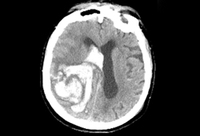

Hemorrhagic stroke
Spot sign on CT angiogram (arrowhead), indicating the presence of hyperdense contrast material within the hematoma bed on postinjection CT; this has been associated with greater risk of subsequent hematoma expansion
Foothills Medical Center personal case files; used with permission
See this image in context in the following section/s:

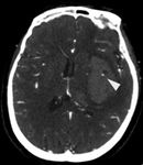
Hemorrhagic stroke
Intracranial hemorrhage on CT scan
Massachusetts General Hospital personal case files; used with permission
See this image in context in the following section/s:

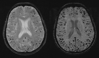
Hemorrhagic stroke
80-year-old woman with numerous punctate foci of hypointensity (black dots) on MRI gradient-echo (GRE) sequence (left), suggesting multiple lobar microbleeds caused by cerebral amyloid angiopathy. MRI susceptibility-weighted imaging (SWI) sequence (right) demonstrates numerous additional microbleeds not seen on the GRE sequence
Foothills Medical Center personal case files; used with permission
See this image in context in the following section/s:
Videos
 Venepuncture and phlebotomy: animated demonstration
How to take a venous blood sample from the antecubital fossa using a vacuum needle.
 How to perform an ECG: animated demonstration
How to record an ECG. Demonstrates placement of chest and limb electrodes.
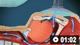
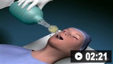 Tracheal intubation: animated demonstration
How to insert a tracheal tube in an adult using a laryngoscope.
 Bag-valve-mask ventilation: animated demonstration
How to use bag-valve-mask apparatus to deliver ventilatory support to adults. Video demonstrates the two-person technique.
Use of this content is subject to our disclaimer