Approach
Diagnosis is based on clinical presentation and a finding of a cardiac mass on echocardiogram, which is typically present in the left atrium. The cardiac mass in the atrium most frequently (in 80% of cases) arises from the region of the septal fossa ovalis.[4] The diagnosis is confirmed by histological examination. The specimen for histology is obtained from either surgery or biopsy via thoracotomy or a transvenous approach.[Figure caption and citation for the preceding image starts]: Large left atrial myxomaFrom the collection of Dr Syed Wamique Yusuf, Department of Cardiology, University of Texas MD Anderson Cancer Center; used with permission [Citation ends].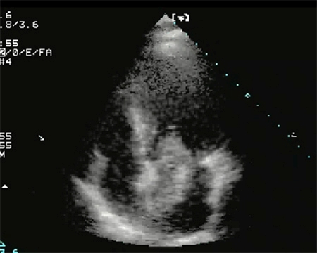 [Figure caption and citation for the preceding image starts]: Two-dimensional echocardiogram showing a right atrial mass suggestive of a myxomaFrom the collection of Dr Syed Wamique Yusuf, Department of Cardiology, University of Texas MD Anderson Cancer Center; used with permission [Citation ends].
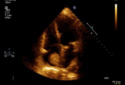 [Figure caption and citation for the preceding image starts]: Atrial myxoma identified on echocardiography (A) and following surgical resection (B)From: HH Ho, WK Seto, E Wang, WH Chow. BMJ Case Reports 2009; doi:10.1136/bcr.2006.093781 [Citation ends].
 [Figure caption and citation for the preceding image starts]: Macroscopic view of a left atrial myxomaR Al-Shahi Salman, D Northridge, ANJ Graham, R Grant. BMJ Case Reports 2009; doi:10.1136/bcr.09.2008.0875 [Citation ends].
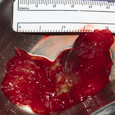
History and examination
Most clinical presentations related to myxoma result from mitral valve obstruction (syncope, dyspnoea, and pulmonary oedema) followed by embolic manifestations.[4][8] Other symptoms include weight loss, fatigue, unexplained fever, and anaemia. Atrial myxoma may result in sudden death. Myxoma involving the right atrium may present with pulmonary embolism. Physical examination may reveal a systolic murmur or a diastolic murmur suggestive of mitral stenosis. A tumour plop may also be heard (a low-pitched diastolic sound as the tumour prolapses into the left ventricle). Texas Heart Institute: heart sounds auscultation Opens in new window[4][8] In one study, the most common auscultation findings were systolic murmur (in 50% of cases) followed by loud first heart sound (32%), an opening snap (26%), and a diastolic murmur (15%).[8] Systolic murmur may be caused by damage to the valves, failure of coaptation of the leaflets, or narrowing of the outflow tract by the tumour. Diastolic murmur is present due to obstruction of the valve caused by the myxoma. Tumour plop may be confused with a mitral opening snap or a third heart sound and can be detected in up to 15% of cases.[4] In one study, a cardiac auscultation abnormality was detected in 64% of patients.[4] Chest examination may reveal fine crepitations consistent with pulmonary oedema.
Extremity examination may also reveal signs of embolic phenomenon. The signs vary depending on the vascular territory involved. Involvement of cerebral vessels results in neurological signs; involvement of coronary arteries may result in an acute myocardial infarction; intestinal obstruction may result in ischaemic bowel; and peripheral arterial obstruction can result in limb-threatening ischaemia.
Imaging
Initial investigations are echocardiogram, ECG, and CXR, and are followed by either a CT or MRI scan. CT and MRI scans provide better delineation of the intracardiac mass, extent of tumour, and extracardiac structures.[10] They also provide anatomical definition for preoperative planning and may also help to identify whether the mass is solid, haemorrhagic, or fatty.[Figure caption and citation for the preceding image starts]: Large left atrial myxomaFrom the collection of Dr Syed Wamique Yusuf, Department of Cardiology, University of Texas MD Anderson Cancer Center; used with permission [Citation ends].[Figure caption and citation for the preceding image starts]: Two-dimensional echocardiogram showing a right atrial mass suggestive of a myxomaFrom the collection of Dr Syed Wamique Yusuf, Department of Cardiology, University of Texas MD Anderson Cancer Center; used with permission [Citation ends].[Figure caption and citation for the preceding image starts]: Chest CT demonstrating a mass in the right atrium (RA) subsequently confirmed to be an atrial myxoma. RV = right ventricle, LV = left ventricleA Yavari, H El-Mahy, ET McWilliams. BMJ Case Reports 2009; doi:10.1136/bcr.10.2008.1031 [Citation ends].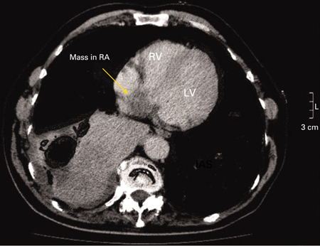
Blood tests
Blood test abnormalities may include:
Anaemia (49% of patients)
Elevated serum gammaglobulin (45% of patients)
Elevated erythrocyte sedimentation rate (55% of patients)
Elevated serum C-reactive protein (75% of patients).[8]
Biopsy
Biopsy is useful in certain conditions, such as differentiating a thrombus from tumour if not already differentiated by echocardiography or MRI/CT scan. It is also helpful in identifying tumours that are unsuitable for surgical resection (e.g., metastatic cardiac tumour) and providing a histological diagnosis, allowing patients to be treated with chemotherapy if appropriate.
Use of this content is subject to our disclaimer