Images and videos
Images


Atrial myxoma
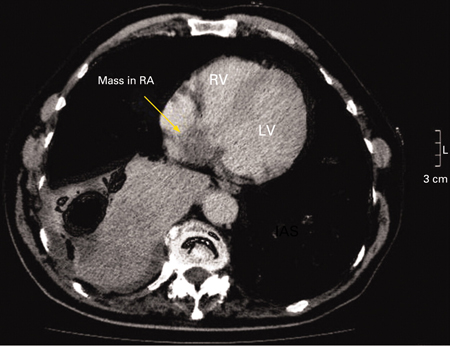
Chest CT demonstrating a mass in the right atrium (RA) subsequently confirmed to be an atrial myxoma. RV = right ventricle, LV = left ventricle
A Yavari, H El-Mahy, ET McWilliams. BMJ Case Reports 2009; doi:10.1136/bcr.10.2008.1031
See this image in context in the following section/s:

Atrial myxoma
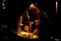
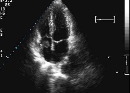
Large left atrial myxoma
From the collection of Dr Syed Wamique Yusuf, Department of Cardiology, University of Texas MD Anderson Cancer Center; used with permission
See this image in context in the following section/s:

Atrial myxoma
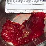
Macroscopic view of a left atrial myxoma
R Al-Shahi Salman, D Northridge, ANJ Graham, R Grant. BMJ Case Reports 2009; doi:10.1136/bcr.09.2008.0875
See this image in context in the following section/s:
Atrial myxoma
Two-dimensional echocardiogram of a right atrial thrombus (note: not attached to septum)
From the collection of Dr Syed Wamique Yusuf, Department of Cardiology, University of Texas MD Anderson Cancer Center; used with permission
See this image in context in the following section/s:

Atrial myxoma
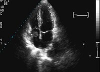
Two-dimensional echocardiogram showing a right atrial mass suggestive of a myxoma
From the collection of Dr Syed Wamique Yusuf, Department of Cardiology, University of Texas MD Anderson Cancer Center; used with permission
See this image in context in the following section/s:
Atrial myxoma
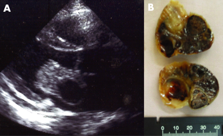
Atrial myxoma identified on echocardiography (A) and following surgical resection (B)
From: HH Ho, WK Seto, E Wang, WH Chow. BMJ Case Reports 2009; doi:10.1136/bcr.2006.093781
See this image in context in the following section/s:
Atrial myxoma
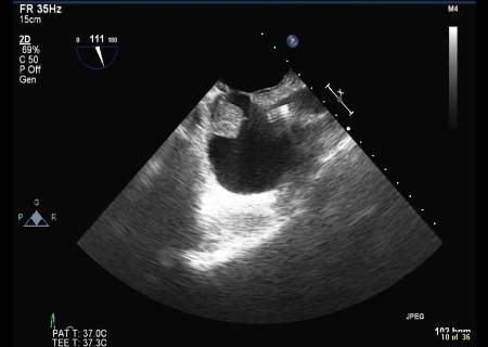
Transoesophageal echocardiogram showing a catheter in the superior vena cava and a right atrial thrombus (note: not attached to septum)
From the collection of Dr Syed Wamique Yusuf, Department of Cardiology, University of Texas MD Anderson Cancer Center; used with permission
See this image in context in the following section/s:
Use of this content is subject to our disclaimer