Images and videos
Images


Haemolytic anaemia
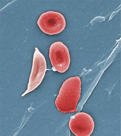
Digitally-colourised scanning electron micrograph showing normal red blood cell (RBCs) and a sickle cell RBC (left) in a blood specimen of patient with sickle cell anaemia
CDC/ Sickle Cell Foundation of Georgia: Jackie George, Beverly Sinclair
See this image in context in the following section/s:
Haemolytic anaemia
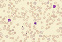
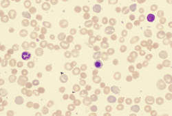
Peripheral blood smear with spherocytes, reticulocytes, and a nucleated red blood cell
From the collection of John Densmore, Department of Medicine, University of Virginia
See this image in context in the following section/s:
Haemolytic anaemia
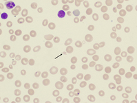
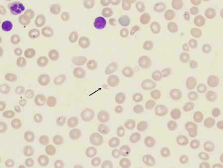
Peripheral blood smear with red blood cell fragments, or schistocytes (arrow)
From the collection of John Densmore, Department of Medicine, University of Virginia
See this image in context in the following section/s:
Haemolytic anaemia
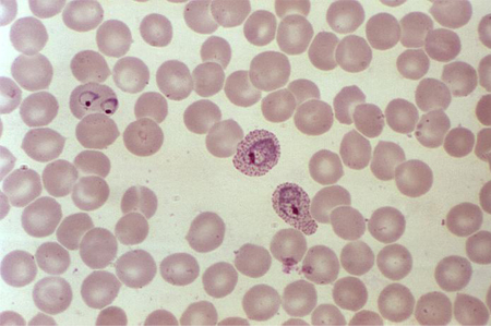
A photomicrograph of a blood smear showing erythrocytes containing developing Plasmodium vivax parasites
CDC/ Dr Mae Melvin
See this image in context in the following section/s:

Haemolytic anaemia
Photomicrograph revealing the presence of what were determined to be numbers of intraerythrocytic Babesia sp. ring-form parasites
CDC/ Dr Mae Melvin
See this image in context in the following section/s:
Videos
 Venepuncture and phlebotomy animated demonstration
How to take a venous blood sample from the antecubital fossa using a vacuum needle.
 Peripheral venous cannulation animated demonstration
How to insert a peripheral venous cannula into the dorsum of the hand.
Use of this content is subject to our disclaimer